🎬 Video Lesson Available
Watch the full 7-slide video lesson for Cell Wall with AI teacher narration and visual explanations.
01Defining the Cell Wall: The Non-Living Protective Boundary of Plant Cells
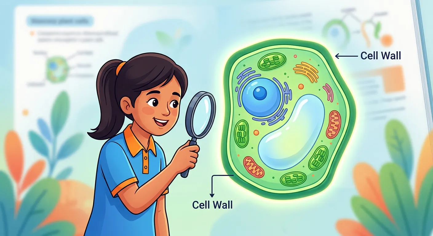
“Welcome, NEET aspirants! Imagine a plant cell as a house in an Indian colony. The cell wall is like the sturdy, protective compound wall surrounding it. It gives the cell its shape, provides mechanical support, and acts as the ultimate security guard for the delicate protoplasm inside.”
In the world of biology, the cell wall stands as a remarkable evolutionary achievement, providing the structural backbone that allows plants to stand tall and thrive in diverse environments. Unlike the fluid and dynamic plasma membrane, the cell wall is a rigid, non-living outer covering that serves as the first line of defense for the delicate protoplasm within. It is not just a passive box but a functional boundary that determines the cell's shape and provides mechanical support against various environmental stresses. For a NEET aspirant, understanding that the cell wall is unique to certain kingdoms—like plants, fungi, and some protists—is fundamental to mastering eukaryotic cell biology.
One of the most critical roles of the cell wall is its protective nature. It acts as a physical barrier that prevents the entry of pathogens and provides resistance against mechanical injury. While the plasma membrane is selectively permeable, regulating the entry and exit of specific solutes, the cell wall is typically quite permeable to water and small solutes, acting more like a protective 'mesh' rather than a solid 'seal'. This distinction is vital for understanding how plants interact with their external environment, especially concerning water uptake and nutrient absorption. Think of the cell wall as the sturdy outer shell of an egg, protecting the life-sustaining yolk and white inside.
Quick Revision Points
- The cell wall is a rigid, non-living structure found outside the plasma membrane.
- Its primary functions include providing shape, mechanical support, and protection from pathogens.
- It acts as a protective shield for the protoplasm, preventing mechanical damage.
- Unlike the plasma membrane, the cell wall is generally permeable to most small molecules.
- It is found in plants, algae, fungi, and bacteria, but is entirely absent in animal cells.
NEET Exam Angle
- Permeability Distinction: Always remember that while the cell wall is permeable, the plasma membrane is selectively permeable. NEET often tests this conceptual difference.
- Non-living Nature: Focus on the fact that the cell wall is a 'secreted' product and is considered non-living, whereas the protoplast is the living component.
- Evolutionary Significance: The presence of a cell wall was a major reason why plants could transition from aquatic to terrestrial environments.
| Feature | Cell Wall | Plasma Membrane |
|---|---|---|
| Nature | Rigid and Non-living | Flexible and Living |
| Permeability | Generally Permeable | Selectively Permeable |
| Primary Function | Protection and Shape | Regulated Transport |
| Composition | Cellulose/Chitin/etc. | Phospholipids and Proteins |
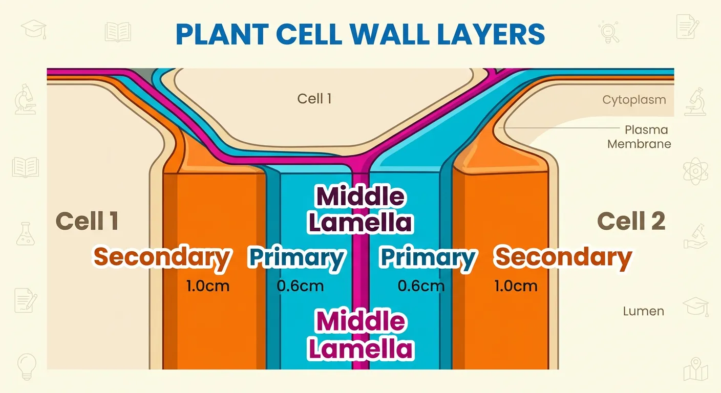
02The Stratified Architecture: Middle Lamella, Primary, and Secondary Walls
“Think of the cell wall like a multi-layered cake! The 'Middle Lamella' is the sticky glue holding neighbors together, made of Calcium pectate. Inside that is the thin, flexible Primary wall, and in mature cells, a thick, rigid Secondary wall develops. It’s all about structural integrity!”
The architecture of the plant cell wall is far from a single, uniform layer. It is a complex, stratified structure composed of distinct layers that develop as the cell grows and matures. The first layer to form is the middle lamella, which acts as the 'cellular glue' that binds adjacent cells together. Chemically, the middle lamella is primarily composed of calcium pectate. This sticky substance ensures that tissues remain cohesive, providing the collective strength required for plant organs like leaves and stems to function as a unit. During the ripening of fruits, this calcium pectate often breaks down, which is why ripe fruits feel soft compared to unripe ones.
Moving inward, we find the primary cell wall. In young, actively growing plant cells, the primary wall is thin, flexible, and capable of extension, allowing the cell to increase in size. As the cell matures and reaches its full size, the growth capability of the primary wall gradually diminishes. In many cells, a secondary wall is then deposited on the inner side of the primary wall, facing the plasma membrane. This secondary wall is much thicker and more rigid, providing the necessary reinforcement for structural cells like tracheids and vessels. It is crucial to note the sequence: the middle lamella is the outermost cementing layer, followed by the primary wall, and then the secondary wall is the innermost layer deposited against the membrane.
Quick Revision Points
- Middle Lamella: Composed of Calcium pectate; cements neighboring cells together.
- Primary Wall: Flexible, capable of growth, found in young cells; thins out as cells mature.
- Secondary Wall: Formed inner to the primary wall; provides structural rigidity in mature cells.
- Developmental Order: Middle Lamella (first) -> Primary Wall -> Secondary Wall (last).
- Pectin's Role: Pectins in the middle lamella are essential for tissue adhesion.
NEET Exam Angle
- Calcium Pectate: This chemical is a high-frequency NEET favorite. Always associate 'Middle Lamella' with 'Calcium Pectate'.
- Position of Secondary Wall: Students often mistake the secondary wall for the outermost layer. Remember: it is formed inside the primary wall.
- Cell Plate Connection: The cell plate formed during cytokinesis eventually matures into the middle lamella.
| Layer | Location | Main Composition | Flexibility |
|---|---|---|---|
| Middle Lamella | Between adjacent cells | Calcium Pectate | Sticky/Adhesive |
| Primary Wall | Outer to Secondary Wall | Cellulose/Hemicellulose | High (Extensible) |
| Secondary Wall | Inner to Primary Wall | Cellulose/Lignin | Low (Rigid) |
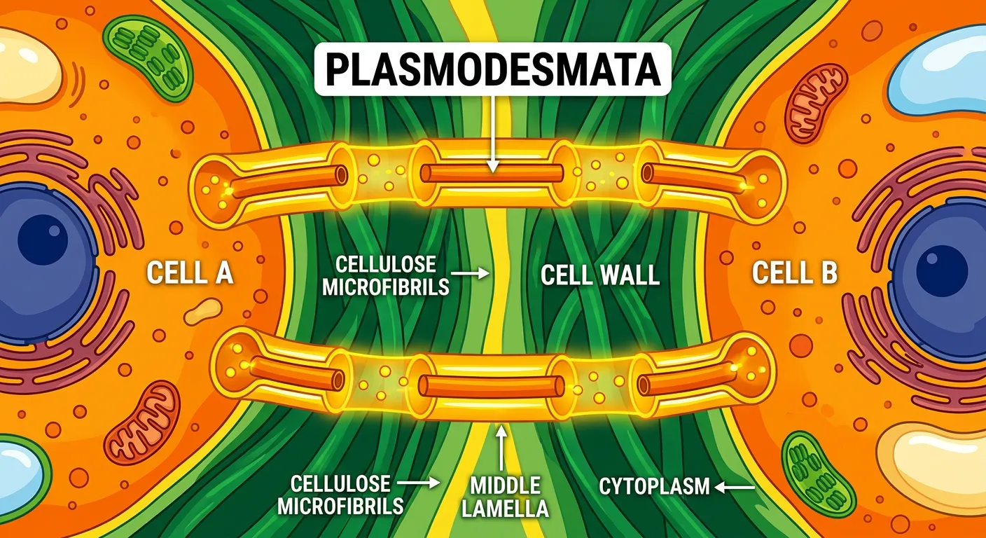
03Plasmodesmata: Cytoplasmic Bridges for Intercellular Communication
“Cells aren't isolated! They have tiny secret tunnels called 'Plasmodesmata' that pierce through the walls. Just like neighbors passing snacks over the fence, these tunnels allow cytoplasm to flow between adjacent cells, helping them communicate and transport nutrients. Biology is all about connections!”
While the cell wall provides a formidable physical barrier, it does not isolate plant cells from one another. Instead, the wall is traversed by microscopic channels known as plasmodesmata. These channels act as cytoplasmic bridges that link the cytoplasm of adjacent cells, creating a continuous network known as the symplast. Through these narrow tunnels, cells can exchange ions, small molecules, and even larger macromolecules like proteins and RNA. This connectivity is essential for coordinating physiological processes across different tissues, allowing the plant to respond as a cohesive organism rather than a collection of independent units.
At the center of most plasmodesmata is a structure called the desmotubule, which is derived from the smooth endoplasmic reticulum (ER). The desmotubule physically connects the ER of one cell to its neighbor, facilitating the direct transport of lipids and other molecules. For NEET aspirants, it is vital to distinguish between the 'apoplastic' pathway—which involves movement through the cell wall and intercellular spaces—and the 'symplastic' pathway, which occurs through the plasmodesmata. The existence of these channels proves that the cell wall is not just a dead boundary, but a sophisticated interface that permits and regulates communication between the living components of the plant.
Quick Revision Points
- Plasmodesmata are cytoplasmic bridges that cross the cell wall and middle lamella.
- They facilitate the movement of nutrients, signals, and water between adjacent cells.
- The Symplast is the system of interconnected protoplasts via plasmodesmata.
- Desmotubules are specialized ER derivatives found within the plasmodesmatal channel.
- They are essential for intercellular communication and metabolic transport in multicellular plants.
NEET Exam Angle
- ER Connection: Recognize the desmotubule as an ER derivative; this is a common point in higher-difficulty MCQ options.
- Transport Pathways: Distinguish between Apoplast (dead) and Symplast (living) pathways. Plasmodesmata are the defining feature of the Symplast.
- Size Exclusion Limit: Understand that only specific molecules can pass through these tunnels, controlled by the 'gating' mechanism of the plasmodesmata.
- Presence: Remember that plasmodesmata are unique to plants and some algae, not found in animal cells (which use gap junctions instead).
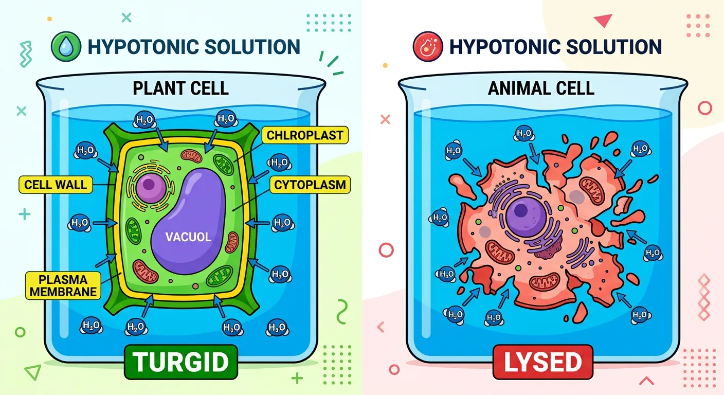
04Turgor Pressure Management: How Cell Walls Prevent Osmotic Lysis
“Why don't plant cells burst like animal cells in water? Because of the cell wall! It exerts 'Turgor Pressure' against the incoming water, keeping the cell firm and upright. It’s like a balloon inside a rigid box—it stays perfectly inflated without popping!”
One of the most fascinating mechanical roles of the cell wall is its ability to manage internal pressure. When a plant cell is placed in a hypotonic solution (like pure water), water enters the cell via osmosis. This causes the protoplast to expand and exert pressure against the rigid cell wall. This internal pressure is known as Turgor Pressure (TP). If a typical animal cell were subjected to such pressure, it would swell and eventually burst (cytolysis). However, the plant cell wall is incredibly strong and exerts an equal and opposite force back onto the protoplast, known as Wall Pressure (WP). This balance keeps the cell firm and turgid.
Turgidity is not just a cellular state; it is the reason why non-woody plants can remain upright. When a plant is well-watered, its cells are turgid, providing a form of 'hydrostatic skeleton' that supports the leaves and stems. Conversely, when a plant loses too much water, turgor pressure drops, the protoplast shrinks away from the wall (plasmolysis), and the plant wilts. Therefore, the cell wall acts as a pressure vessel that allows the plant to maintain its posture and survive in varying osmotic conditions. For NEET students, this interaction between the cell wall and the vacuole is a key concept in plant physiology.
Quick Revision Points
- Turgor Pressure (TP): Pressure exerted by the protoplast against the cell wall.
- Wall Pressure (WP): Pressure exerted by the cell wall against the protoplast.
- Cytolysis Prevention: The rigid cell wall prevents the cell from bursting in hypotonic environments.
- Turgidity: Essential for maintaining the upright posture of herbaceous plants.
- Plasmolysis: The process where the protoplast shrinks away from the cell wall due to water loss.
NEET Exam Angle
- Hypotonic Environment: Remember that plant cells thrive in hypotonic conditions because the cell wall allows them to become turgid without bursting.
- Formulaic Relationship: At full turgidity, TP = WP. This equilibrium is crucial for osmotic calculations in the Plant Physiology unit.
- Animal vs. Plant: A common NEET question asks why plant cells don't burst in water while RBCs do. The answer is always the presence of the cell wall.
| Condition | Plant Cell Behavior | Animal Cell Behavior |
|---|---|---|
| Hypotonic | Becomes Turgid (Safe) | Swells and Bursts (Lysis) |
| Isotonic | Becomes Flaccid | Remains Normal |
| Hypertonic | Plasmolyzed | Shriveled (Crenated) |
05Chemical Composition: The Tensile Strength of Cellulose Microfibrils
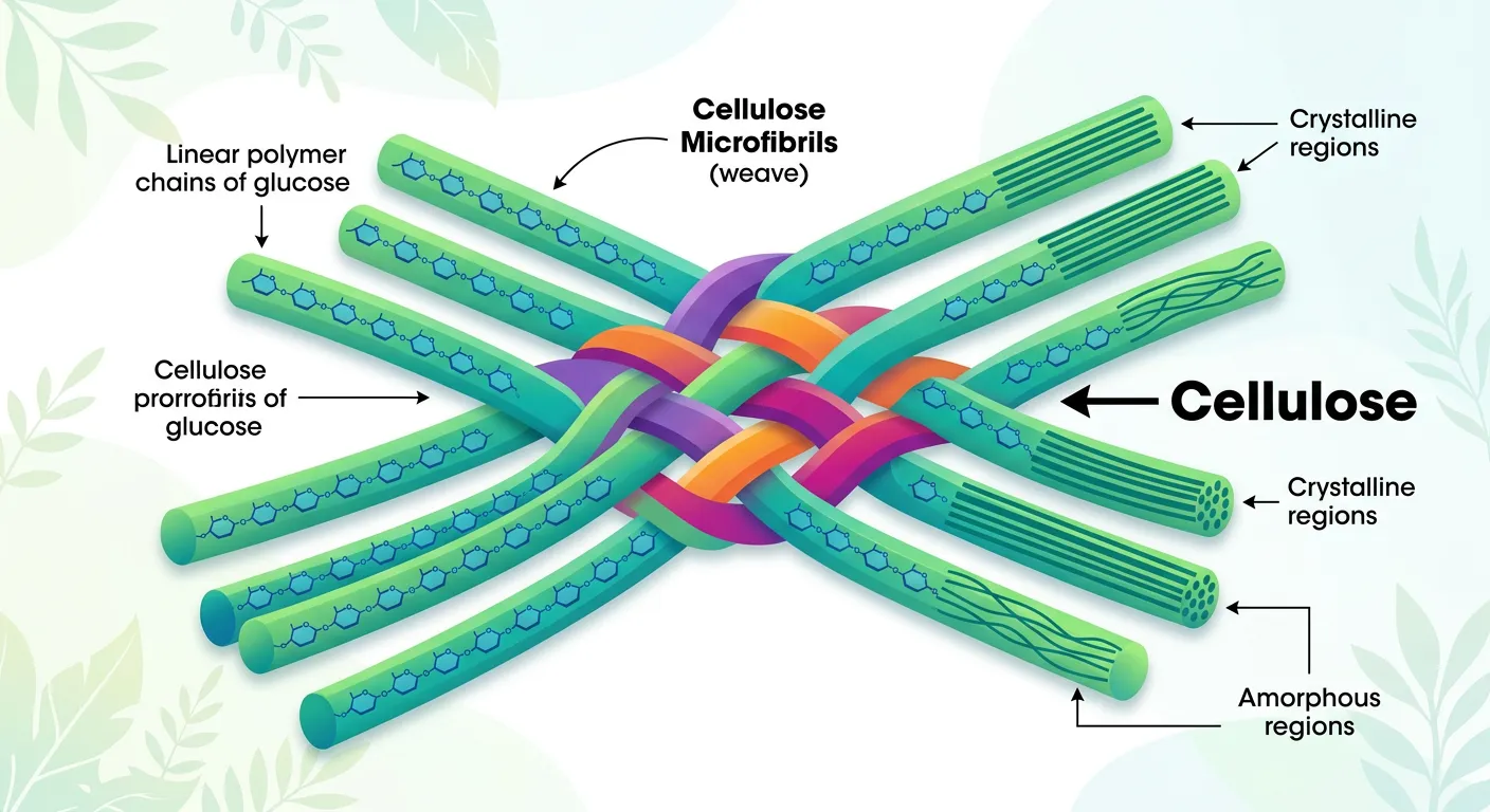
“What is this wall made of? Mostly cellulose! Think of cellulose as long, strong, fibrous ropes woven together to create a mesh. This gives the plant cell its incredible tensile strength, allowing trees to grow tall and withstand harsh winds without falling over.”
The incredible strength of the cell wall is a result of its unique chemical composition, dominated by the polysaccharide cellulose. Cellulose is a linear polymer of beta-glucose units, which bundle together to form long, fibrous structures called microfibrils. These microfibrils act like the steel rebar in reinforced concrete, providing immense tensile strength. In the primary wall, these microfibrils are often arranged in a loose, mesh-like fashion, which allows for cellular expansion. In the secondary wall, however, they are deposited in thick, parallel layers, often oriented in different directions to maximize rigidity and resistance to compression.
Beyond cellulose, the cell wall matrix is filled with other complex carbohydrates like hemicellulose and pectin. Hemicellulose binds the cellulose microfibrils together, while pectin forms a hydrated gel that fills the gaps, giving the wall its semi-solid consistency. In some specialized tissues, additional substances like lignin, suberin, or cutin are deposited to provide waterproofing or extra mechanical reinforcement. For instance, lignin is what makes wood 'woody' and extremely hard. Understanding this chemistry is vital for NEET, as it connects cell biology to the larger unit of biomolecules and plant anatomy.
Quick Revision Points
- Cellulose: The primary structural component; a polymer of beta-D-glucose.
- Microfibrils: Bundles of cellulose chains that provide high tensile strength.
- Hemicellulose: A cross-linking polymer that ties microfibrils together.
- Pectin: A matrix polysaccharide that determines the wall's porosity and charge.
- Tensile Strength: Allows plants to resist being pulled or snapped by wind and gravity.
NEET Exam Angle
- Biomolecule Connection: Be ready to identify cellulose as a homopolymer of beta-glucose. This often bridges the Cell and Biomolecules chapters.
- Tensile Strength: Understand that the arrangement of microfibrils is what gives the wall its specific mechanical properties.
- Matrix Components: While cellulose is the 'fiber', remember that hemicellulose and pectin are the 'matrix' or 'filler'.
- Abundance: Cellulose is the most abundant organic polymer on Earth—a fun fact that occasionally appears in ecological contexts.
06NEET Comparative Focus: Algae, Fungi, and Higher Plant Variations
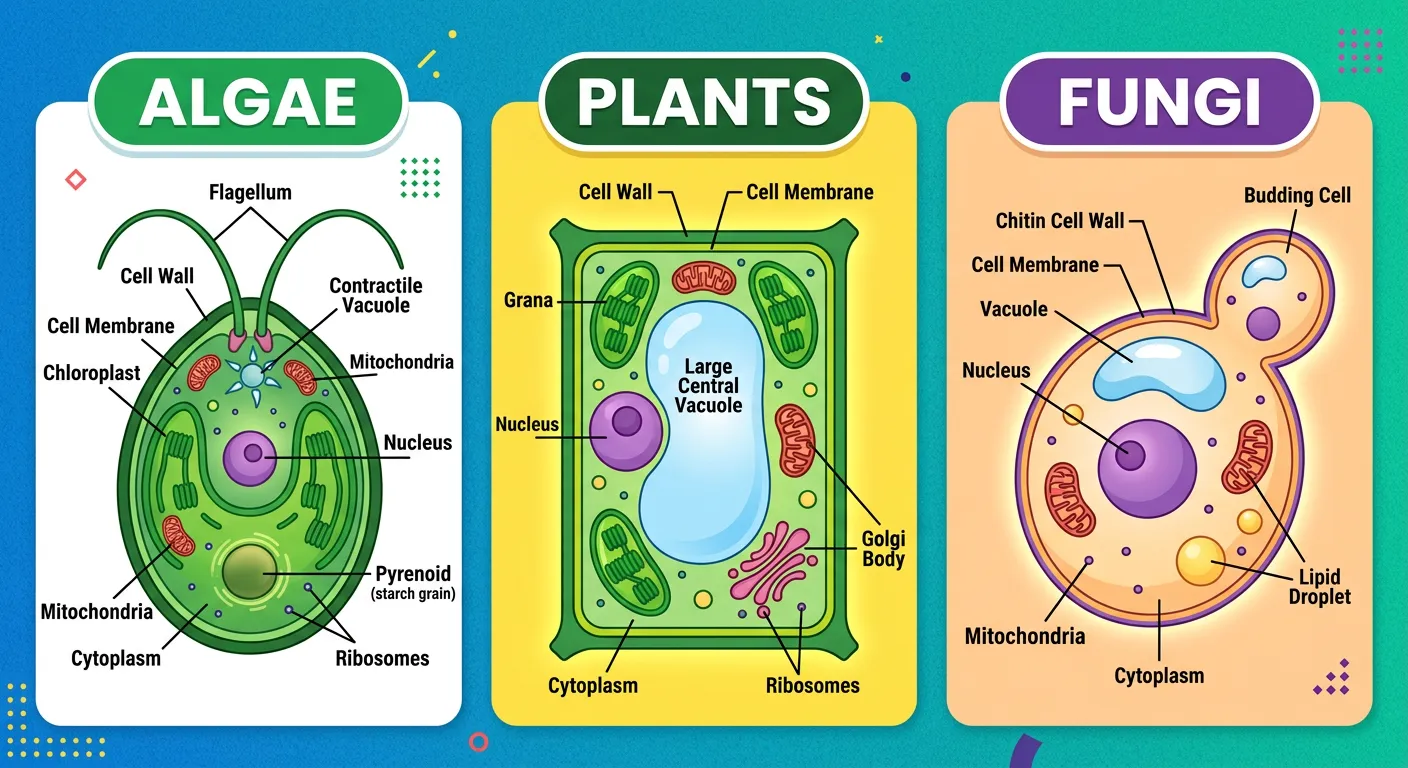
“NEET trap alert! Not all cell walls are the same. Algae have cellulose, galactans, and minerals. Plants are cellulose-heavy. But Fungi? They go for Chitin! Always remember the material: it’s a favorite question in competitive exams. Know your chemistry, know your biology!”
A common 'trap' in the NEET biology paper involves the varying composition of cell walls across different organisms. While we often generalize that cell walls are made of cellulose, this is only strictly true for higher plants. Algae, for example, have a cell wall composed of cellulose, but it also contains unique components like galactans, mannans, and minerals such as calcium carbonate. This distinct chemistry helps algae survive in aquatic environments where osmotic pressures and physical stresses differ from those on land. Recognizing these specific algal components is a high-yield area for competitive exams.
Fungi represent another major variation. The fungal cell wall is primarily composed of chitin, a tough, nitrogen-containing polysaccharide. Chitin is actually a polymer of N-acetylglucosamine (NAG) and is much more resistant to chemical breakdown than cellulose. This is a crucial adaptation for fungi, many of which are decomposers or pathogens. Bacteria, though not usually the primary focus of the eukaryotic cell wall topic, have walls made of peptidoglycan (murein). As a medical aspirant, you must be able to quickly distinguish these materials, as they are the targets of various antifungal and antibiotic treatments.
Quick Revision Points
- Algae: Cellulose + Galactans + Mannans + Calcium Carbonate.
- Higher Plants: Predominantly Cellulose + Hemicellulose + Pectin.
- Fungi: Chitin (Polymer of N-acetylglucosamine).
- Bacteria: Peptidoglycan (Murein).
- Evolutionary Note: Chemical differences in cell walls reflect the different ecological niches occupied by these kingdoms.
NEET Exam Angle
- Algal Components: NEET has specifically asked about 'galactans and mannans' in the context of algal cell walls multiple times (e.g., NEET 2020).
- Chitin: Always remember that fungi do NOT have cellulose walls. Chitin is their signature molecule.
- N-acetylglucosamine (NAG): Know that this is the monomer of chitin for inter-disciplinary questions involving Biomolecules.
| Organism Group | Primary Component | Key Additional Substances |
|---|---|---|
| Green Plants | Cellulose | Hemicellulose, Pectin |
| Algae | Cellulose | Galactans, Mannans, CaCO3 |
| Fungi | Chitin | Glucans, Proteins |
| Bacteria | Peptidoglycan | Teichoic acids (in Gram +) |
07Conclusion: The Cell Wall as a Dynamic and Living Shield
“So, we've covered the structure, the layers, the connections, and the composition of the cell wall. It’s not just a boundary; it’s a complex, living shield! Keep revising these core concepts, and you’ll be ready to crack that NEET paper with ease. Happy studying!”
To conclude our deep dive into the cell wall, it is essential to move past the idea that it is merely a static, 'dead' box. While the wall itself is non-living, it is a dynamic shield that is constantly being modified by the living protoplast within. From the sticky calcium pectate of the middle lamella to the extensible primary wall and the reinforced secondary wall, every layer has a specific developmental and functional purpose. The cell wall integrates perfectly with the internal physiology of the cell, managing turgor pressure and facilitating communication through plasmodesmata to ensure the plant’s survival and growth.
For your NEET preparation, the strategy should be to focus on the 'odd-one-out' facts: the specific minerals in algae, the chemical nature of the middle lamella, and the inward deposition of the secondary wall. These details are what distinguish a top-ranking student from the rest. The cell wall is a perfect example of how structure meets function in biology—its rigidity providing the strength for trees to reach the sky, while its microscopic pores keep the lines of communication open. Keep these core concepts in mind, visualize the layered 'cake' structure, and you will find this topic to be one of the most scoring sections in your biology syllabus.
Quick Revision Points
- The cell wall is a complex, multi-layered structure (Middle Lamella, Primary, Secondary).
- It provides mechanical support, shape, and protection from biotic and abiotic stress.
- Plasmodesmata ensure that the cell is not isolated, but part of a symplastic network.
- The composition varies strictly by kingdom (Cellulose in plants vs. Chitin in fungi).
- Turgor pressure management is the wall's key role in plant osmoregulation.
NEET Exam Angle
- Final Summary: Focus on the 'Middle Lamella' as the first-formed layer and its chemical (Calcium pectate).
- Memory Hook: Use 'C-H-P' for Plant walls (Cellulose, Hemicellulose, Pectin) and 'C-G-M' for Algal walls (Cellulose, Galactans, Mannans).
- Question Pattern: Most questions involve identifying the correct components for a specific group or the correct sequence of wall layers.
- Revision Strategy: Review the diagram of a plant cell wall at least thrice before the exam to lock in the spatial arrangement of layers.
Recommended Reading
Explore related Biology topics to build deeper chapter connections for NEET.
- Cell Theory · Topic 3.1
- Golgi Bodies · Topic 3.10
- Lysosomes · Topic 3.11
- Vacuoles · Topic 3.12
- Plastids · Topic 3.15
- Prokaryotic and Eukaryotic Cell · Topic 3.2
- Jump to Key Terms (Quick Revision)
- Review Common NEET Mistakes
- Read Topic FAQs
- Check PYQ Pattern Notes
- Practice NEET MCQs
- Solve NEET PYQs
📚 Key Terms
⚠️ Common NEET Mistakes
- 1Thinking the secondary wall is formed on the outside of the primary wall; it is actually deposited on the inner side (towards the membrane).
- 2Confusing the cell wall (non-living and permeable) with the plasma membrane (living and selectively permeable).
- 3Assuming all eukaryotic cell walls are made of cellulose, forgetting that fungi use chitin and algae have galactans/mannans.
- 4Forgetting that animal cells never have a cell wall, which is a major distinguishing feature in classification questions.
- 5Ignoring the role of Calcium pectate in the middle lamella, which is a frequent target for specific chemical-based MCQs.
📝 NEET PYQ Pattern
NEET frequently asks about the chemical composition of algal cell walls (Cellulose, Galactans, Mannans) and the specific material of the Middle Lamella (Calcium pectate). Questions on the distinction between primary and secondary walls (position and growth capability) and the chitinous nature of fungal walls are also very common (e.g., NEET 2016, 2019, and 2021).
❓ Frequently Asked Questions
What is the chemical composition of the middle lamella in plant cells?
The middle lamella is primarily composed of Calcium pectate. It acts as a cementing layer that glues adjacent plant cells together.
How does the cell wall composition of algae differ from higher plants?
While higher plants mainly have cellulose, hemicellulose, and pectin, algal cell walls contain cellulose along with galactans, mannans, and minerals like calcium carbonate.
What is the primary function of plasmodesmata in multicellular plants?
Plasmodesmata serve as cytoplasmic bridges between adjacent cells, allowing for the transport of water, nutrients, and signaling molecules, thereby connecting the individual protoplasts into a symplastic network.
Why does the primary cell wall diminish as the cell matures?
The primary cell wall is flexible and capable of growth. As the cell reaches maturity and stops expanding, the growth potential of the primary wall decreases, and a more rigid secondary wall is often deposited inner to it.
What substance makes up the cell wall of fungi, and how does it differ from plants?
Fungal cell walls are made of chitin (a polymer of N-acetylglucosamine), whereas plant cell walls are primarily made of cellulose (a polymer of beta-glucose).
How does the cell wall help a plant cell survive in a hypotonic medium?
In a hypotonic medium, water enters the cell, creating turgor pressure. The rigid cell wall exerts an equal and opposite wall pressure, which prevents the cell membrane from over-expanding and bursting (cytolysis).
Written By
NEET Content Strategist & Biology Expert
Sangita Kumari is a NEET educator and content strategist with over 6 years of experience teaching Biology, Chemistry, and Physics to Class 11 and 12 aspirants. She helps bridge the gap between traditional NCERT preparation and modern AI-powered learning. Her content is trusted by thousands of NEET aspirants across India.