🎬 Video Lesson Available
Watch the full 7-slide video lesson for Golgi Bodies with AI teacher narration and visual explanations.
01The Discovery and Molecular Logistics of the Golgi Apparatus
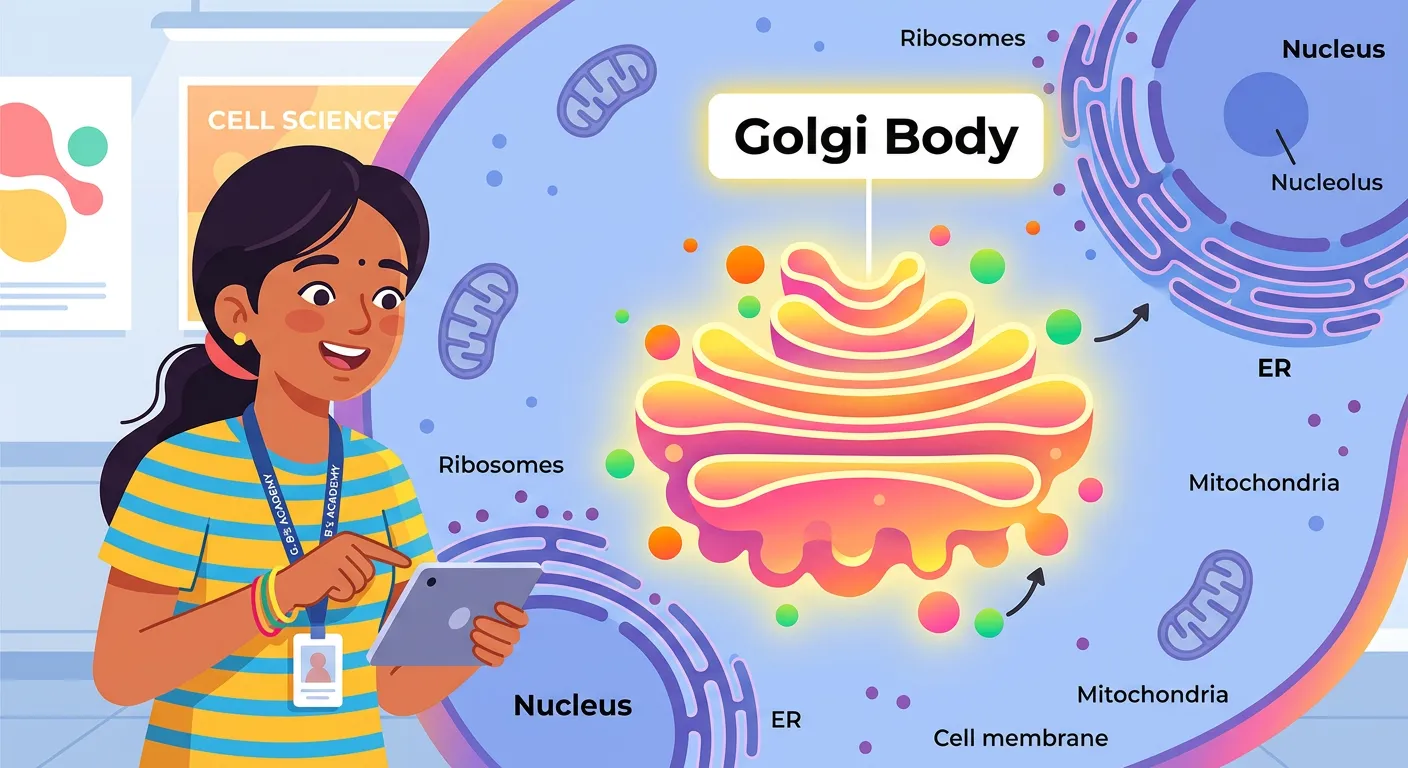
“Meet the Golgi Body! Think of it as the 'Amazon fulfillment center' of your cell. Discovered by Camillo Golgi, this stack of flattened sacs is the ultimate processing hub. Every protein needs a final check before it goes live, and the Golgi is the master organizer.”
In the complex architectural landscape of the eukaryotic cell, few organelles possess a history as rich as the Golgi body. It was first identified in 1898 by the Italian cytologist Camillo Golgi. Using a specific silver staining technique known as the 'black reaction' (la reazione nera), he observed a reticular (net-like) structure located near the nucleus of nerve cells. This discovery was groundbreaking, as it unveiled a previously invisible layer of cellular organization. For NEET aspirants, it is essential to remember that this organelle is an integral part of the endomembrane system, working in tight coordination with the Endoplasmic Reticulum (ER) and Lysosomes.
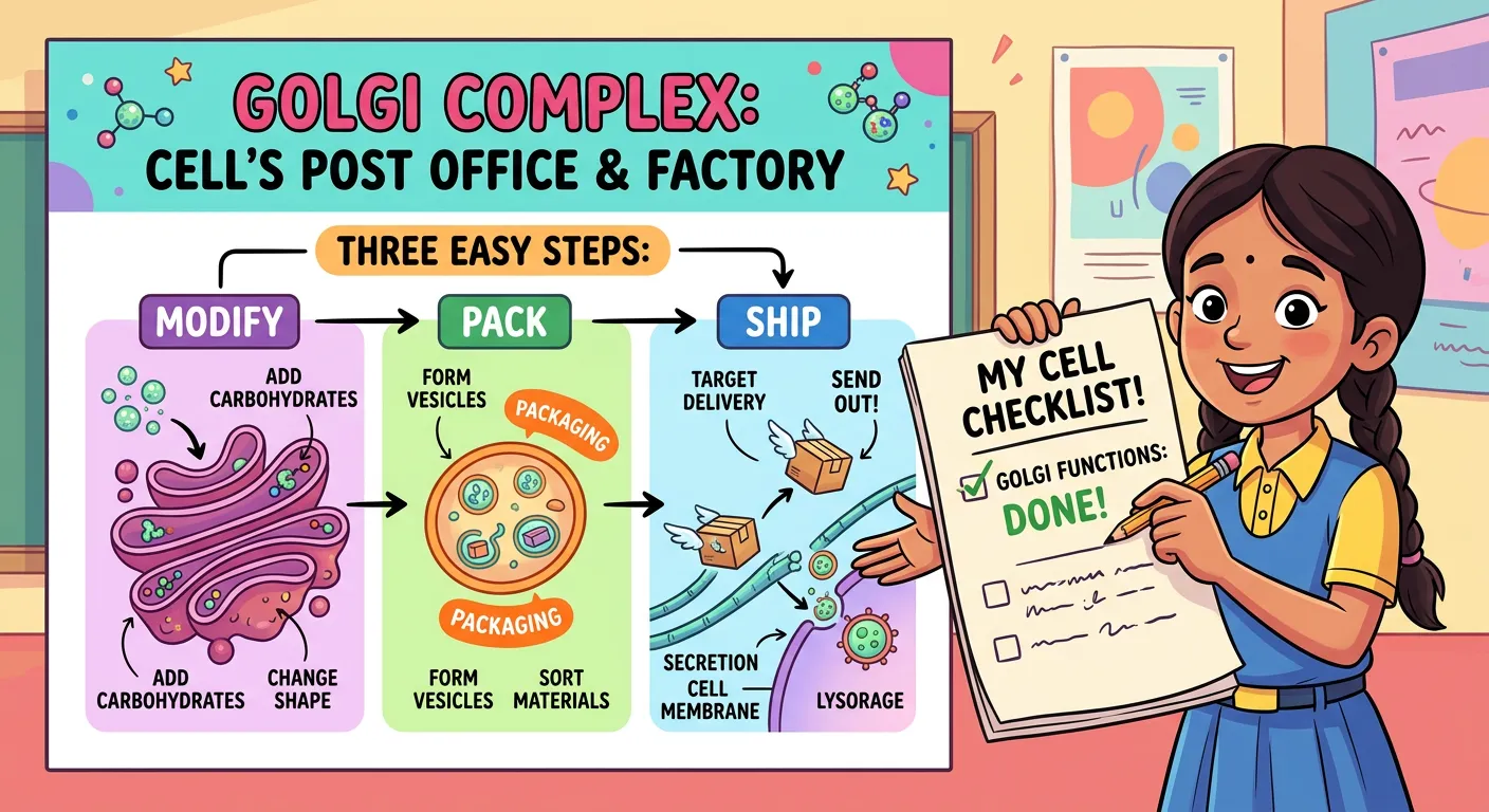
To understand the Golgi apparatus practically, imagine it as a high-tech 'Amazon Fulfillment Center' or a central post office. While the Endoplasmic Reticulum functions like a factory producing raw goods (proteins and lipids), these goods cannot be shipped out immediately. They are 'unfinished' and lack specific destination tags. The Golgi body steps in to receive these raw materials, inspect them for quality, modify them biochemically, and finally package them into neat vesicles for delivery. Without this logistics hub, the cell would suffer from molecular chaos, with proteins ending up in the wrong organelles or failing to reach the cell exterior.
Physically, the Golgi complex consists of many flat, disc-shaped sacs or cisternae of 0.5µm to 1.0µm diameter. These are stacked parallel to each other like a pile of coins or pancakes. While the number of cisternae varies depending on the cell's metabolic activity—being highly abundant in secretory cells like those in the pancreas—the fundamental principle remains the same: it is the master organizer of cellular traffic.
Quick Revision Points
- Discovery: Camillo Golgi (1898) using silver staining in nerve cells.
- Analogy: The 'Post Office' or 'Packaging Center' of the cell.
- Component: Consists of flattened sacs called cisternae (0.5–1.0 µm).
- System: Part of the endomembrane system, ensuring coordinated functioning.
- Location: Typically situated near the nucleus in animal cells.
NEET Exam Angle
- Historical Fact: Questions often ask for the scientist and the year (Camillo Golgi, 1898).
- Staining Technique: The use of silver salts for visualization is a recurring MCQ point.
- Cell Type: Remember that Golgi bodies are absent in mature mammalian RBCs and prokaryotes.
| Feature | Details for NEET |
|---|---|
| Discovered By | Camillo Golgi (1898) |
| Staining Method | Silver Staining (Black Reaction) |
| Structural Unit | Cisternae (Flattened sacs) |
| Coordination | Works with ER and Lysosomes |
02Structural Polarity: Navigating the Cis and Trans Faces
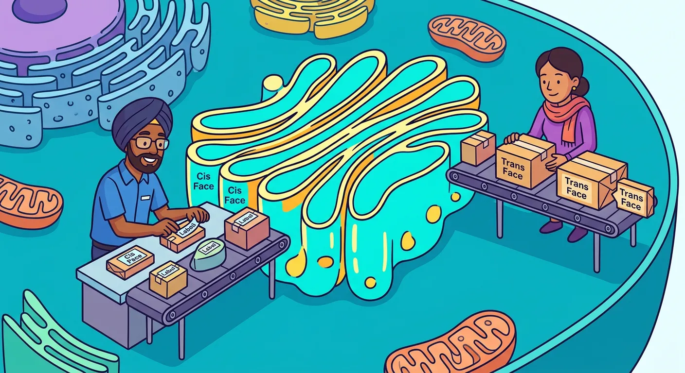
“The Golgi has two distinct faces: the Cis face, which acts as the 'receiving dock' near the ER, and the Trans face, which is the 'shipping dock' facing the plasma membrane. Proteins arrive, get sorted, and are ready for their final delivery across the cell.”
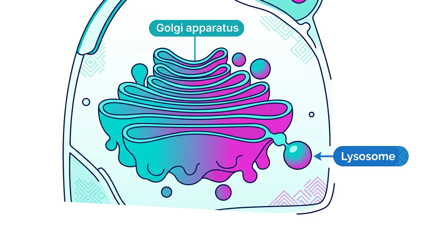
The Golgi apparatus is not just a random pile of sacs; it possesses a highly defined structural and functional polarity. This 'sidedness' is crucial for the unidirectional flow of materials. The Golgi has two distinct faces: the 'Cis' face and the 'Trans' face. The Cis face, also known as the 'forming face,' is convex in shape and is positioned close to the Rough Endoplasmic Reticulum (RER). This serves as the receiving dock where transition vesicles, carrying proteins from the ER, merge with the Golgi membrane to dump their cargo.
On the opposite side lies the Trans face, also called the 'maturing face.' This face is concave and oriented toward the plasma membrane. It acts as the shipping dock of the cell. Once proteins have travelled through the various layers of cisternae and undergone modification, they are pinched off from the Trans face as secretory vesicles. It is vital to note that while the Cis and Trans faces are interconnected and work together, they are biochemically distinct, containing different enzymes suited for specific stages of protein processing.
This polarity ensures that the cell maintains a 'one-way street' for molecular traffic. A common point of confusion for students is the physical relationship between the ER and the Golgi. Because the Golgi modifies what the ER produces, it must stay in close proximity. This spatial arrangement is why you will always find the forming face (Cis) nestled near the nuclear envelope and ER, while the maturing face (Trans) is primed to release vesicles toward the cell periphery or other organelles.
Quick Revision Points
- Cis Face: Forming face, convex shape, faces the Endoplasmic Reticulum.
- Trans Face: Maturing face, concave shape, faces the plasma membrane.
- Polarity: The Golgi is a polarized organelle with a distinct receiving and shipping side.
- Vesicle Flow: Materials move from ER → Cis Face → Cisternae → Trans Face → Destination.
- Function: This arrangement allows for sequential modification of proteins.
NEET Exam Angle
- Face Identification: NEET frequently tests the difference between the convex/forming (Cis) and concave/maturing (Trans) faces.
- Proximity: Why is Golgi near ER? To receive vesicles (transition vesicles) effectively.
- Unidirectional Flow: Understand that proteins do not move backward from Trans to Cis under normal conditions.
| Feature | Cis Face (Forming) | Trans Face (Maturing) |
|---|---|---|
| Shape | Convex | Concave |
| Orientation | Towards Nucleus/ER | Towards Plasma Membrane |
| Function | Receiving vesicles from ER | Releasing secretory vesicles |
| NEET Keyword | Forming Face | Maturing Face |
03Glycosylation: The Golgi's Role in Protein Modification
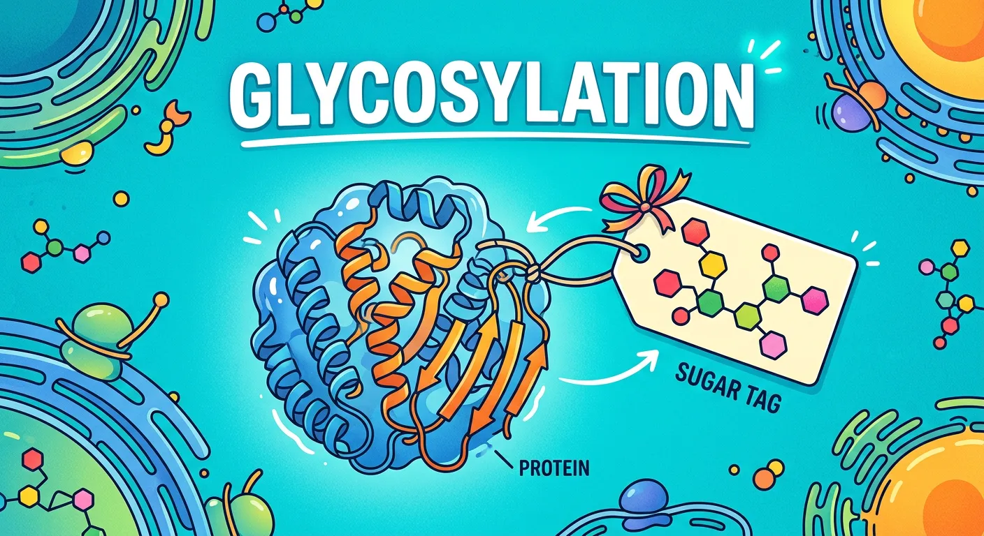
“Ever added a topping to your favorite street food? That's what the Golgi does! Through 'Glycosylation', it adds sugar molecules to proteins. These tags tell the protein where to go—like a courier address—ensuring they reach the right destination without any errors.”
If the Endoplasmic Reticulum is the factory that assembles the 'basic' protein chain, the Golgi apparatus is the specialized workshop that adds the 'finish' and 'branding.' The most critical biochemical function of the Golgi is Glycosylation. This is the process of adding carbohydrate moieties (sugar molecules) to proteins and lipids to form glycoproteins and glycolipids. These sugar chains act as molecular 'barcodes' or 'zip codes,' telling the cell exactly where the molecule belongs—whether it should go to the lysosome, be embedded in the cell membrane, or be secreted outside the cell.
For NEET aspirants, it is crucial to distinguish between N-linked and O-linked glycosylation. While N-linked glycosylation usually begins in the RER and is refined in the Golgi, O-linked glycosylation (where sugars are attached to the oxygen atom of Serine or Threonine residues) occurs almost exclusively in the Golgi complex. This modification is essential for the stability and functionality of proteins. For instance, many of the proteins on the surface of your cells that determine your blood type are actually glycoproteins modified by the Golgi.
Beyond glycosylation, the Golgi is involved in 'signal tagging.' Specific enzymes within the cisternae add phosphate groups or other chemical tags to proteins. This ensures high-fidelity trafficking. If a protein is destined for the lysosome, it receives a specific 'Mannose-6-Phosphate' tag. Without these modifications, the cell would be unable to distinguish between a digestive enzyme and a structural protein, leading to internal degradation and cellular death.
Quick Revision Points
- Major Site: The Golgi is the primary site for the synthesis of glycoproteins and glycolipids.
- Glycosylation: Adding sugar chains to proteins (glycoproteins) and lipids (glycolipids).
- O-linked Glycosylation: Specifically occurs in the Golgi apparatus.
- Molecular Tags: Chemical modifications serve as addresses for protein delivery.
- Enzymatic Hub: Contains various glycosyltransferases for sugar addition.
NEET Exam Angle
- High-Yield Question: "Which organelle is responsible for the formation of glycoproteins and glycolipids?" Answer: Golgi apparatus.
- Biomolecule Link: Connect this to the 'Biomolecules' chapter regarding the structure of complex carbohydrates.
- Tagging: Understand the concept of signal sequences and tags (like M6P) for organelle targeting.
| Modification Type | Resulting Molecule | Primary Location |
|---|---|---|
| Glycosylation of Proteins | Glycoproteins | Golgi (Major site) |
| Glycosylation of Lipids | Glycolipids | Golgi |
| Phosphorylation | Activated Enzymes | Golgi Cisternae |
| Sulfation | Sulfated Proteoglycans | Trans-Golgi Network |
04Secretory Vesicles and the Cellular Export Mechanism
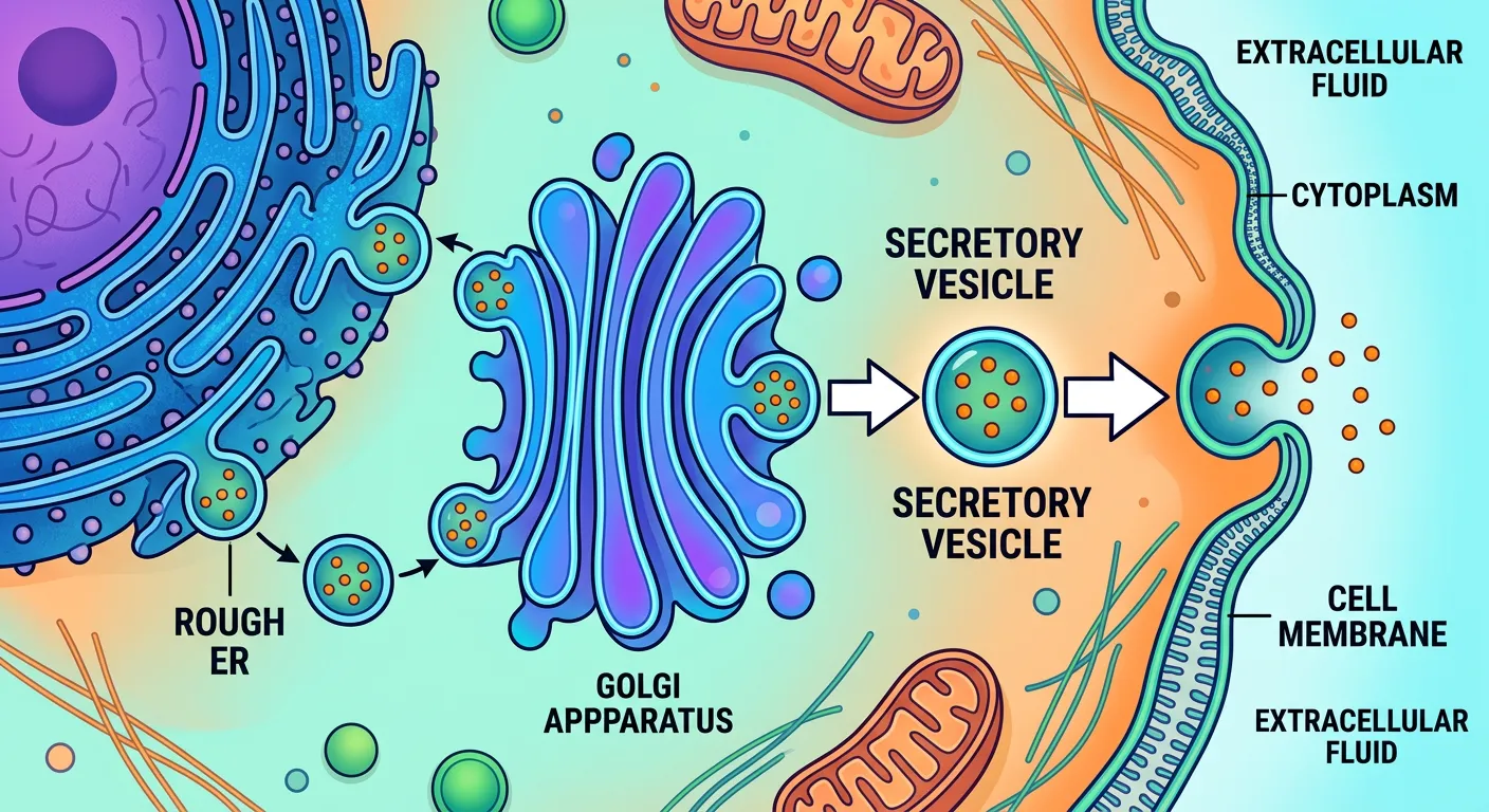
“Once the cargo is processed and tagged, it gets packed into 'Secretory Vesicles'. These tiny bubbles pinch off from the Trans face, acting like mini-couriers that carry the finished products either to the cell surface for export or to other organelles that need supplies.”
Once the Golgi has finished modifying and tagging its cargo, it must prepare the products for dispatch. This happens at the Trans-Golgi Network (TGN), the final exit point of the organelle. Here, the membrane of the Trans face buds outward and pinches off to form tiny, membrane-bound spheres called 'Secretory Vesicles.' These vesicles are like the delivery vans of the cell, loaded with enzymes, hormones, or neurotransmitters that need to be sent to specific locations.
There are two main pathways for these vesicles: the intracellular pathway and the extracellular pathway. In the intracellular pathway, vesicles might carry digestive enzymes to a lysosome. In the extracellular (secretory) pathway, the vesicles move toward the plasma membrane. Through a process called exocytosis, the vesicle membrane fuses with the cell's outer membrane, and the contents are spilled into the external environment. This is how your pancreas releases insulin into your bloodstream or how nerve cells release chemical signals.
An often-overlooked function of this process is membrane recycling. As secretory vesicles fuse with the plasma membrane, they add new lipids and proteins to the cell surface. This helps maintain the surface area of the cell, which might otherwise shrink due to endocytosis. Thus, the Golgi is not just a packaging center; it is a vital player in maintaining the dynamic integrity of the cell's boundary. For students, understanding this bulk transport mechanism is key to mastering both 'Cell Structure' and 'Transport in Plants/Humans.'
Quick Revision Points
- Vesicle Budding: Process where the Trans face membrane pinches off to form carriers.
- Exocytosis: Fusion of secretory vesicles with the plasma membrane to release cargo.
- Cargo Types: Includes hormones, mucus, digestive enzymes, and cell wall materials.
- Membrane Flow: Golgi contributes to the growth and repair of the plasma membrane.
- Directionality: Moves from the Trans-Golgi Network to the target site.
NEET Exam Angle
- Exocytosis: Know that this is an active process requiring energy and membrane fusion.
- Secretory Cells: Cells rich in Golgi (like glandular cells) have high rates of vesicle formation.
- Flow sequence: Always remember the sequence: RER → Transition Vesicle → Cis Golgi → Trans Golgi → Secretory Vesicle → Plasma Membrane.
05Lysosome Biogenesis: The Golgi as a Producer of Recycling Centers
“Did you know the Golgi is a parent too? It plays a crucial role in forming Lysosomes—the cell’s recycling centers. It packages digestive enzymes into these vesicles, ensuring the cell can break down waste effectively. It truly is the busiest organelle in town!”
One of the most fascinating 'parental' roles of the Golgi apparatus is the production of lysosomes. Lysosomes are the 'suicide bags' or recycling centers of the cell, filled with powerful hydrolytic enzymes (acid hydrolases). These enzymes are originally synthesized by ribosomes on the Rough ER and then transported to the Golgi. The Golgi then takes these enzymes, concentrates them, and packages them into specialized vesicles that eventually become 'primary lysosomes.'
This functional relationship is encapsulated in the term 'GERL Complex'—which stands for Golgi-Endoplasmic Reticulum-Lysosome. This complex describes how these three organelles work in a synchronized pipeline to manage intracellular digestion and waste disposal. The Golgi's role here is highly specialized: it must ensure that these powerful enzymes are sequestered safely within a membrane. If these enzymes were released freely into the cytoplasm, they would digest the cell itself (autolysis).
To prevent self-digestion during the packaging process, the Golgi adds specific carbohydrate markers to the lysosomal enzymes. The most famous of these is the Mannose-6-Phosphate (M6P) tag. Receptors in the Trans-Golgi membrane recognize this tag and gather all M6P-labeled enzymes into a single budding vesicle. This precision is a testament to the Golgi's role as a master regulator of cellular health. Without the Golgi's packaging expertise, the cell would lack its primary defense against invading pathogens and damaged organelles.
Quick Revision Points
- Origin of Lysosomes: Primary lysosomes bud off from the Trans face of the Golgi.
- GERL Complex: Integrated system of Golgi, ER, and Lysosomes for waste management.
- Enzyme Packaging: Acid hydrolases are concentrated and membrane-bound by the Golgi.
- M6P Tag: Mannose-6-Phosphate is the specific signal for lysosomal targeting.
- Protection: Packaging prevents enzymes from damaging the host cell cytoplasm.
NEET Exam Angle
- GERL Concept: Be ready for questions on which organelles constitute the GERL system.
- Hydrolytic Enzymes: Remember these enzymes are synthesized in RER but packaged in Golgi.
- Primary vs. Secondary: The Golgi produces the primary lysosome (inactive enzymes).
| Organelle | Primary Role in GERL |
|---|---|
| Rough ER | Synthesis of hydrolytic enzymes |
| Golgi Body | Processing and packaging of enzymes into vesicles |
| Lysosome | Intracellular digestion and waste breakdown |
06Dictyosomes: Understanding the Golgi Architecture in Plants
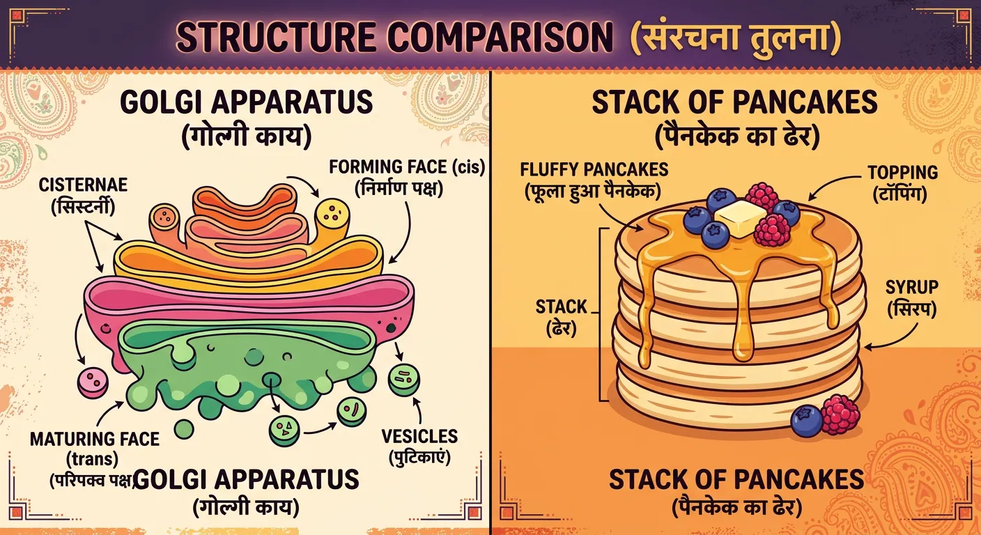
“In botany, we call these stacks 'Dictyosomes'. Imagine a stack of fluffy pancakes or flat pita bread—that’s exactly how the cisternae are arranged. This unique structure provides maximum surface area for the chemical modifications needed to keep your cellular functions running smoothly.”
While the Golgi apparatus is a universal eukaryotic organelle, it presents itself with a fascinating twist in the plant kingdom. In animal cells, the Golgi usually appears as a single, large, interconnected complex—often called a 'Golgi ribbon'—located near the nucleus. However, in plant cells, the organelle is broken up into many smaller, scattered units known as 'Dictyosomes.' These dictyosomes are distributed throughout the cytoplasm, and unlike their animal counterparts, they function as independent modular units rather than a single centralized hub. This decentralized arrangement is likely an adaptation to the large central vacuole found in plants, which pushes the cytoplasm and its organelles toward the cell periphery.
The classic 'Pancake Stack' morphology is highly evident in dictyosomes, where each stack typically contains 4 to 8 cisternae. This arrangement provides a massive surface area for essential chemical reactions. In plants, the Golgi has a very specific and critical job: the synthesis of non-cellulosic cell wall polysaccharides. While cellulose itself is synthesized by enzyme complexes located directly at the plasma membrane, the Golgi (Dictyosome) is the primary factory for hemicellulose and pectin. These complex carbohydrates are then transported via vesicles to the growing cell wall to provide structural support.
Furthermore, during plant cell division (mitosis), these dictyosomes play a leading role. They migrate to the center of the dividing cell to help form the 'phragmoplast' and subsequently the 'cell plate.' This cell plate acts as the precursor to the new cell wall that will eventually divide the two daughter cells. This function is a frequent subject of NEET questions, as it highlights the organelle's role in cytokinesis. Understanding dictyosomes is essential for aspirants because it demonstrates how organelles adapt to specific metabolic needs. Because plants are constantly building and reinforcing their cell walls, they require many small 'local' units spread throughout the cell. This allows for the rapid and efficient delivery of building materials to multiple points simultaneously, ensuring structural integrity during rapid growth and development.
Quick Revision Points
- Dictyosomes: The term used for Golgi bodies specifically in plant cells.
- Morphology: Occur as multiple, independent stacks of cisternae scattered in the cytoplasm.
- Plant Function: Major site for synthesis of cell wall pectin and hemicellulose.
- Cell Division: Involved in the formation of the 'cell plate' during cytokinesis.
- Surface Area: The stacked structure maximizes the area for enzymatic activity.
NEET Exam Angle
- Terminology: Always associate 'Dictyosomes' with plant cells.
- Cell Wall Synthesis: A common MCQ asks which organelle synthesizes pectin/hemicellulose.
- Cytokinesis: Note the Golgi's role in phragmoplast and cell plate formation in plants.
07NEET Exam Focus: Summary of the Endomembrane System
“Let’s recap for your NEET exam: The Golgi body is for packaging, modifying, and shipping proteins. Remember, it’s the postmaster of the cell! Master this, and you’ve aced a key part of the endomembrane system. Stay curious, keep studying, and let’s crush those exams!”
As we conclude our deep dive into the Golgi body, let’s synthesize everything for the NEET exam. The Golgi apparatus does not exist in isolation; it is the 'middle man' of the endomembrane system. The sequence of events is always: Synthesis (ER) → Modification & Packaging (Golgi) → Delivery (Vesicles). If you master this flow, you can answer almost any question regarding cellular logistics. Remember the key term: Glycosylation. If you see 'glycoprotein' or 'glycolipid' in a question, your mind should immediately go to the Golgi.
From a structural perspective, remember the polarity. The convex 'Cis' face receives and the concave 'Trans' face releases. This polarity is a common trap in exams. Also, don't forget the specialized roles: forming the acrosome of sperm (essential for fertilization) and producing the lysosomes that keep the cell clean. In plants, think 'Dictyosomes' and 'Cell Wall.' These are the high-yield associations that separate top scorers from the rest.
Finally, visualize the Golgi as the postmaster. It checks the address (tags), puts the items in boxes (vesicles), and sends them out. It ensures that the cell remains a functional, organized unit rather than a bag of mixed chemicals. Keep this mental model in mind as you review the other organelles of the endomembrane system—the ER, Vacuoles, and Lysosomes—as they all function as part of this one big, beautiful, coordinated molecular machine.
Quick Revision Points
- Sequence: Nucleus → RER → Cis Golgi → Trans Golgi → Plasma Membrane.
- Key Synthesis: Glycoproteins and Glycolipids.
- Plant Equivalent: Dictyosomes (scattered units).
- Vital Roles: Acrosome formation in sperm, lysosome biogenesis, cell plate formation.
- Exam Tip: Focus on the 'polarity' and 'packaging' functions.
NEET Exam Angle
- Integration: Expect questions that link the Golgi to the ER and Lysosomes (Endomembrane system).
- High-Yield Fact: The Golgi is the primary site for the formation of the acrosome in animal sperm cells.
- PYQ Focus: Site of glycosylation and the receiving/shipping face orientation.
| Function | Biological Significance |
|---|---|
| Packaging | Prevents enzymatic damage to the cell |
| Glycosylation | Vital for cell-cell recognition and signaling |
| Acrosome Formation | Necessary for sperm to penetrate the egg |
| Pectin Synthesis | Essential for plant cell wall strength |
Recommended Reading
Explore related Biology topics to build deeper chapter connections for NEET.
- Cell Theory · Topic 3.1
- Lysosomes · Topic 3.11
- Vacuoles · Topic 3.12
- Plastids · Topic 3.15
- Prokaryotic and Eukaryotic Cell · Topic 3.2
- Plant and Animal Cell · Topic 3.3
- Jump to Key Terms (Quick Revision)
- Review Common NEET Mistakes
- Read Topic FAQs
- Check PYQ Pattern Notes
- Practice NEET MCQs
- Solve NEET PYQs
📚 Key Terms
⚠️ Common NEET Mistakes
- 1Confusing the Cis face (forming) with the Trans face (maturing) in terms of shape and orientation.
- 2Thinking that cellulose is synthesized in the Golgi; while pectin and hemicellulose are Golgi-derived, cellulose is made at the plasma membrane.
- 3Assuming Golgi bodies are present in all eukaryotic cells (they are absent in mature mammalian RBCs and sieve tubes).
- 4Believing that the Golgi synthesizes proteins; it only modifies and packages them (synthesis occurs in the ER/ribosomes).
- 5Underestimating the polarity of the organelle—forgetting that materials move in a specific one-way direction.
📝 NEET PYQ Pattern
Recent NEET trends (2018–2024) show a heavy focus on the Golgi apparatus as the 'site of synthesis of glycoproteins and glycolipids.' Questions also frequently test the structural polarity (Cis vs Trans faces) and the organelle's role in the endomembrane system. Students should also be prepared for questions linking the Golgi to the acrosome of sperm and cell plate formation in plants.
❓ Frequently Asked Questions
Which organelle is responsible for the formation of glycoproteins and glycolipids?
The Golgi apparatus is the primary site for the synthesis of glycoproteins and glycolipids through a process called glycosylation.
What is the functional difference between the cis and trans faces of the Golgi apparatus?
The cis face (forming face) is convex and receives transition vesicles from the Endoplasmic Reticulum, while the trans face (maturing face) is concave and releases secretory vesicles destined for the cell surface or other organelles.
Why is the Golgi apparatus called the 'post office' or 'packaging center' of the cell?
It is called the post office because it receives proteins from the ER, sorts them, adds molecular 'address tags' (like sugar chains), and packages them into vesicles for delivery to specific destinations.
Are Golgi bodies present in prokaryotic cells? Explain.
No, Golgi bodies are membrane-bound organelles and are therefore absent in prokaryotic cells (like bacteria). They are also absent in mature mammalian red blood cells (RBCs).
What is the relationship between the Golgi body and the Endoplasmic Reticulum?
The Golgi and ER work closely together as part of the endomembrane system. The ER synthesizes proteins and lipids, which are then sent in vesicles to the Golgi's cis face for modification and final packaging.
What are Dictyosomes, and how do they differ from animal Golgi bodies?
Dictyosomes are the subunits of the Golgi apparatus found in plant cells. Unlike the single, large interconnected Golgi complex in animal cells, dictyosomes are many small, scattered stacks of cisternae distributed throughout the plant cytoplasm.
Written By
NEET Content Strategist & Biology Expert
Sangita Kumari is a NEET educator and content strategist with over 6 years of experience teaching Biology, Chemistry, and Physics to Class 11 and 12 aspirants. She helps bridge the gap between traditional NCERT preparation and modern AI-powered learning. Her content is trusted by thousands of NEET aspirants across India.