🎬 Video Lesson Available
Watch the full 7-slide video lesson for Cell Organelles with AI teacher narration and visual explanations.
01The Selective Gateway: Cell Membrane Dynamics and Permeability

“Imagine your cell is a bustling Indian smart city! The Cell Membrane is like the city's high-tech security gate. It decides exactly who gets to enter or leave, keeping the city safe and organized. This 'selective permeability' is the first secret to life itself!”
The cell membrane, or plasma membrane, is far more than just a physical boundary; it is a sophisticated, semi-permeable filter that dictates the internal environment of the cell. In 1972, Singer and Nicolson proposed the 'Fluid Mosaic Model,' which remains the most widely accepted explanation for its structure. Imagine a fluid 'sea' of phospholipids where proteins float like icebergs. This lipid bilayer is composed of amphipathic molecules—having both a hydrophilic (water-loving) polar head and a hydrophobic (water-fearing) non-polar tail. The arrangement ensures that the non-polar tails are protected from the aqueous environment, maintaining the membrane's structural integrity.
Proteins within this mosaic are categorized based on their ease of extraction: peripheral proteins lie on the surface, while integral proteins are partially or totally buried in the membrane. These proteins serve as channels, carriers, and receptors. One of the most critical features of the membrane is its fluidity, which is essential for functions like cell growth, formation of intercellular junctions, secretion, and endocytosis. Cholesterol, tucked between the fatty acid tails, acts as a 'temperature buffer,' preventing the membrane from becoming too rigid in the cold or too fluid in the heat.
When we talk about transport, the membrane is 'selectively permeable.' This means it allows some molecules like neutral solutes to pass via simple diffusion according to the concentration gradient, while others, like polar molecules, require carrier proteins. Water moves by osmosis. However, moving ions or molecules against their concentration gradient (from low to high concentration) requires energy in the form of ATP. This is known as active transport, perfectly exemplified by the Sodium-Potassium (Na+/K+) pump found in nerve cells.
Quick Revision Points
- Fluid Mosaic Model: Proposed by Singer and Nicolson (1972); emphasizes the quasi-fluid nature of lipids.
- Lipid Composition: Primarily phosphoglycerides with polar heads facing outwards and hydrophobic tails facing inwards.
- Protein Types: Integral (transmembrane) and Peripheral (surface-associated).
- Transport Mechanisms: Passive (no ATP) vs. Active (uses ATP, e.g., Na+/K+ pump).
- Carbohydrates: Often present as glycoproteins or glycolipids for cell-to-cell recognition.
NEET Exam Angle
- Frequent Question: Identification of the 'fluid' part (lipids) vs the 'mosaic' part (proteins).
- Membrane Ratios: Remember that the ratio of protein to lipid varies; in human erythrocytes (RBCs), it is approximately 52% protein and 40% lipids.
- Polarity: Understand that polar molecules cannot pass through the non-polar lipid bilayer without carrier proteins.
| Transport Type | Energy Required | Direction of Flow | Example |
|---|---|---|---|
| Simple Diffusion | No | Along Gradient | Oxygen/CO2 |
| Facilitated Diffusion | No | Along Gradient (via Protein) | Glucose via GLUT |
| Active Transport | Yes (ATP) | Against Gradient | Na+/K+ Pump |
02The Nucleus: Genomic Governance and Cellular Command
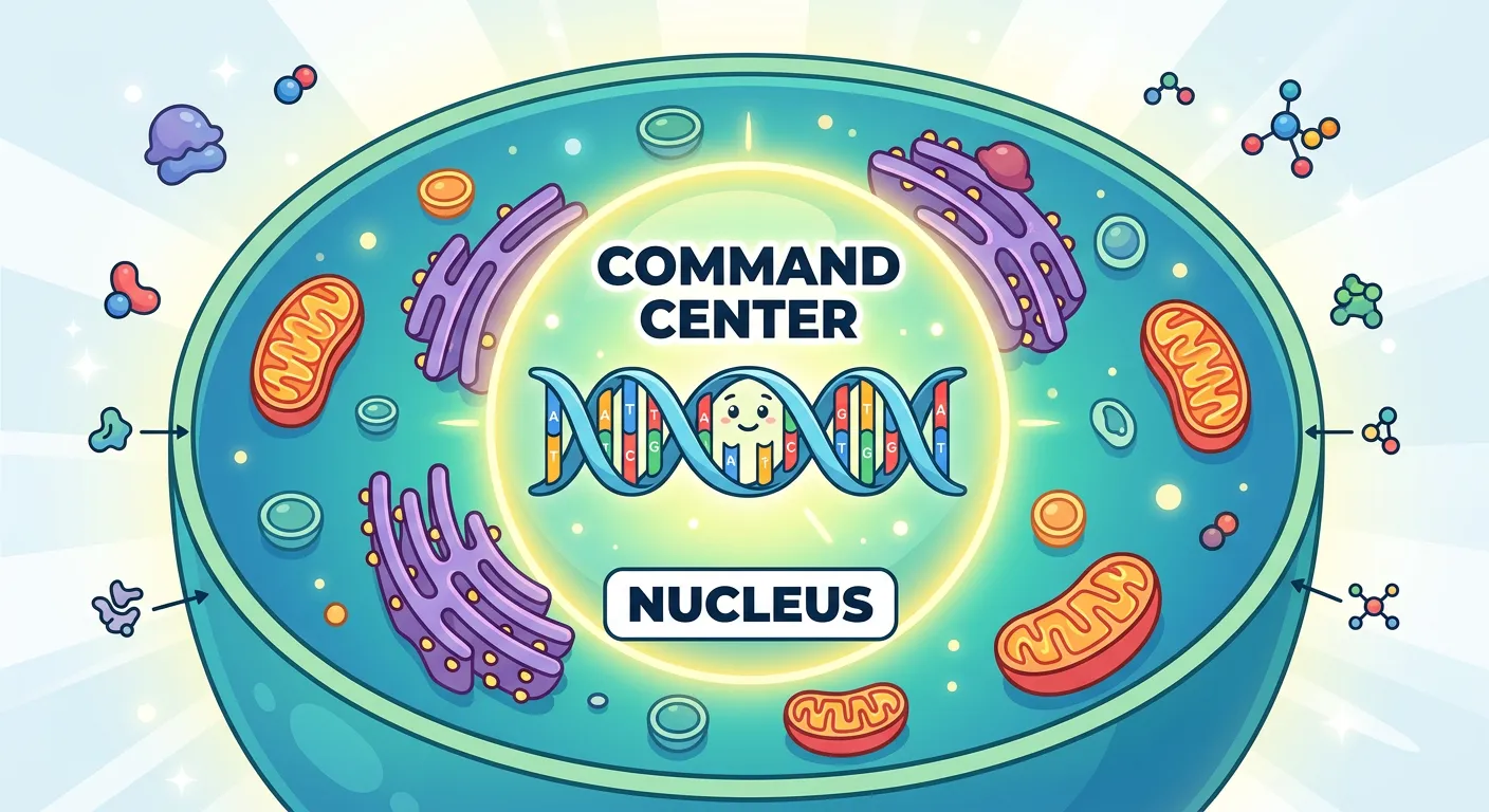
“Meet the Nucleus, the 'Prime Minister's Office' of the cell. It houses the DNA, which is our master instruction manual. Every growth, repair, or function is commanded from here. Think of it as the ultimate brain that holds the blueprints for your entire biological identity.”
The nucleus, first described by Robert Brown in 1831, is the control center of the eukaryotic cell. It contains the majority of the cell's genetic material, organized as multiple extremely long linear DNA molecules in complex with a large variety of proteins, such as histones, to form chromosomes. During the interphase (the non-dividing stage), the nucleus contains a loose network of nucleoprotein fibers called chromatin. However, during various stages of cell division, cells show structured chromosomes in place of the nucleus.
The nucleus is bounded by a double-membrane structure called the nuclear envelope. The space between these two membranes (10 to 50 nm) is called the perinuclear space, which acts as a barrier between the materials inside the nucleus and the cytoplasm. The outer membrane usually remains continuous with the endoplasmic reticulum and also bears ribosomes. At several places, the nuclear envelope is interrupted by minute pores, which are formed by the fusion of its two membranes. These nuclear pores provide passages for the movement of RNA and protein molecules in both directions between the nucleus and the cytoplasm.
Inside the nucleus, we find the nucleoplasm, which contains the nucleolus and chromatin. The nucleolus is a spherical, non-membrane bound structure that is continuous with the rest of the nucleoplasm. It is the primary site for active ribosomal RNA (rRNA) synthesis. Cells that are actively involved in protein synthesis usually have larger and more numerous nucleoli. Understanding the organization of the nucleus is fundamental for mastering genetics and molecular biology topics later in the NEET syllabus.
Quick Revision Points
- Discovery: Robert Brown (1831); term 'Chromatin' coined by Flemming.
- Nuclear Envelope: Double membrane with perinuclear space and nuclear pores.
- Nucleolus: Site for rRNA synthesis; not membrane-bound.
- Chromatin: Contains DNA, basic proteins (histones), non-histone proteins, and RNA.
- Nuclear Pores: Regulate bidirectional traffic of RNA and proteins.
NEET Exam Angle
- Histones: Note that histones are 'basic' proteins (rich in lysine and arginine), a fact often tested in Molecular Basis of Inheritance.
- Nucleolus Function: Always associate the nucleolus with 'protein synthesis machinery' or 'ribosome factory.'
- Chromosomes: Remember that a single human cell has approximately 2 meters of DNA distributed among its 46 chromosomes.
03Mitochondria: ATP Production and the Endosymbiotic Theory
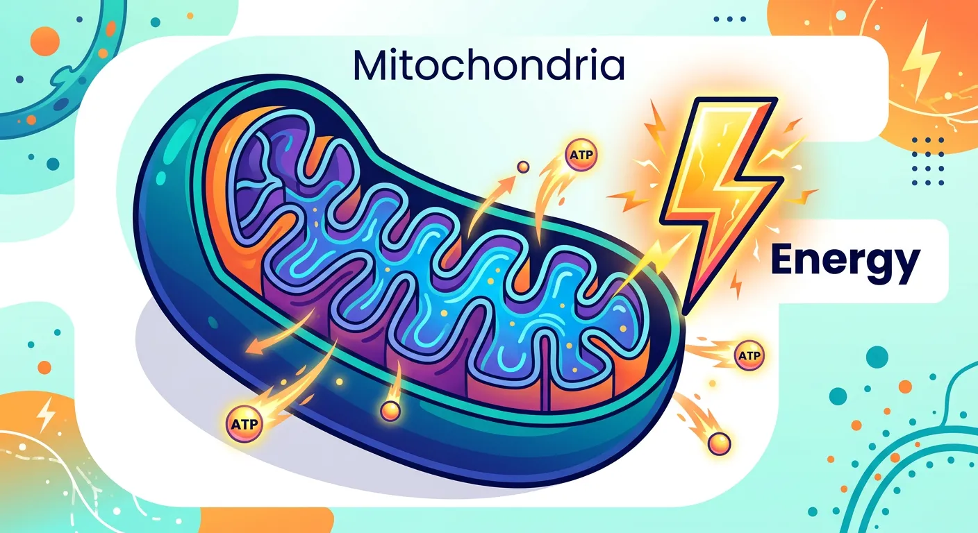
“Ever wondered where your energy for long study nights comes from? Meet the Mitochondria, the 'Powerhouse of the Cell'! It burns glucose like a power plant to produce ATP. Without these cellular batteries, you wouldn't have the energy to even blink or study for your NEET exam!”
Often referred to as the 'powerhouse of the cell,' mitochondria are double-membrane-bound organelles responsible for aerobic respiration and ATP (Adenosine Triphosphate) production. They are generally sausage-shaped or cylindrical, with a diameter of 0.2–1.0 μm. The number of mitochondria per cell varies significantly depending on the metabolic activity of the tissue. For instance, muscle cells and the flight muscles of birds contain thousands of mitochondria, while less active cells contain fewer.
The structure consists of an outer membrane and an inner membrane, which divides its lumen into two aqueous compartments: the outer compartment and the inner compartment (the matrix). The inner membrane forms numerous infoldings called cristae that project into the matrix. These cristae are vital because they significantly increase the surface area available for the Electron Transport System (ETS) and ATP synthase enzymes. The outer membrane serves as a limiting boundary and contains porins that make it quite permeable to small molecules.
A fascinating aspect of mitochondria is their semi-autonomous nature. They possess their own circular DNA molecule, a few RNA molecules, and 70S ribosomes—similar to those found in bacteria. This supports the 'Endosymbiotic Theory,' which suggests mitochondria were once free-living prokaryotes that entered into a symbiotic relationship with early eukaryotic cells. Because they can synthesize some of their own proteins and divide by fission, they are not entirely dependent on the nuclear genome.
Quick Revision Points
- Structure: Double membrane; inner membrane forms cristae to increase surface area.
- Matrix: Contains enzymes for the Krebs Cycle (TCA cycle).
- Powerhouse: Site of aerobic respiration; produces ATP ('Energy Currency').
- Semi-autonomous: Has own circular DNA and 70S ribosomes.
- Reproduction: Divides by fission, similar to bacteria.
NEET Exam Angle
- Ribosome Type: Be careful—mitochondria have 70S ribosomes, while the cytoplasm has 80S. This is a common trap in MCQ options.
- Membrane Characteristics: The inner membrane is highly impermeable compared to the outer membrane.
- DNA Shape: Always remember mitochondrial DNA is circular, not linear.
| Feature | Outer Membrane | Inner Membrane |
|---|---|---|
| Permeability | Highly permeable (porins) | Selectively permeable |
| Folds | Smooth/Continuous | Folded into Cristae |
| Enzyme Content | Involved in lipid synthesis | Involved in ETS and ATP synthesis |
| Surface Area | Lower | Higher (due to cristae) |
04Endoplasmic Reticulum: The Manufacturing and Logistics Network
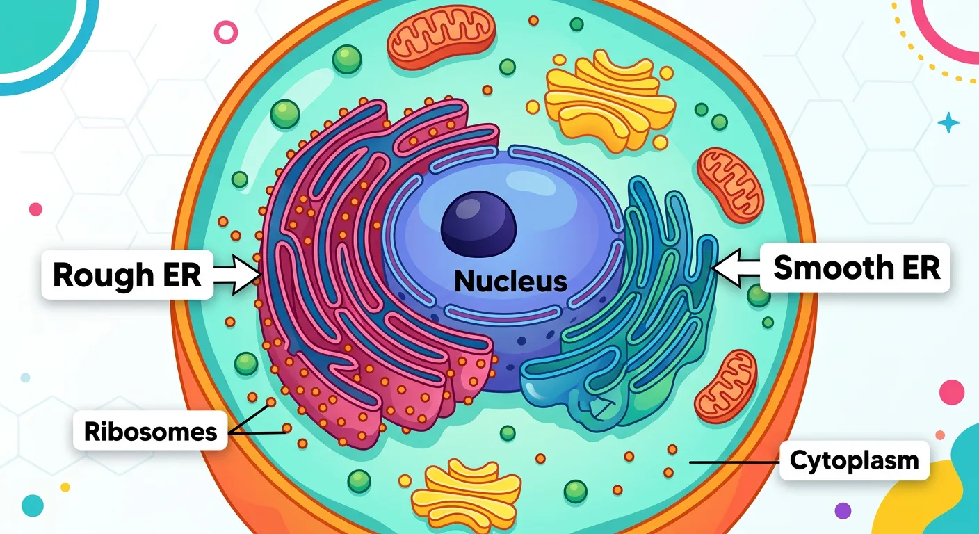
“The Endoplasmic Reticulum is our cell's manufacturing hub. The 'Rough' side is covered in ribosomes, acting like a protein factory assembly line, while the 'Smooth' side builds lipids. It’s like a massive logistics department that packages and ships essential materials throughout the cell's busy streets.”
The Endoplasmic Reticulum (ER) is an extensive network of tiny tubular structures scattered in the cytoplasm, dividing the intracellular space into two distinct compartments: luminal (inside ER) and extra-luminal (cytoplasm). The ER is physically continuous with the outer nuclear membrane and extends throughout the cell. It exists in two morphologically and functionally distinct forms: Rough Endoplasmic Reticulum (RER) and Smooth Endoplasmic Reticulum (SER).
RER is characterized by the presence of ribosomes on its outer surface. Because ribosomes are the sites of protein synthesis, RER is heavily involved in the synthesis and secretion of proteins. Cells actively involved in protein secretion, such as pancreatic cells producing digestive enzymes, have extensive RER. The RER is mainly composed of flattened sac-like structures called cisternae. In contrast, the SER lacks ribosomes and appears smooth. It is the major site for the synthesis of lipids and lipid-like steroidal hormones (e.g., estrogen, testosterone). In liver cells, SER plays a vital role in detoxifying various drugs and poisons.
The ER also acts as an intracellular transport system, moving materials between the nucleus and the cytoplasm or between different parts of the cytoplasm. In muscle cells, a specialized form of SER called the sarcoplasmic reticulum stores calcium ions (Ca2+), which are essential for muscle contraction—a crucial point of connection between cell biology and human physiology.
Quick Revision Points
- RER: Studded with ribosomes; primary site for protein synthesis and secretion.
- SER: No ribosomes; primary site for lipid/steroid synthesis and detoxification.
- Structure: Network of cisternae (RER), tubules, and vesicles (SER).
- Compartmentalization: Divides cytoplasm into luminal and extra-luminal spaces.
- Continuity: Outer nuclear membrane is usually continuous with RER.
NEET Exam Angle
- Cell Type Examples: Expect questions asking which organelle is abundant in fibroblasts (RER) vs. sebaceous glands (SER).
- Detoxification: SER's role in the liver is a frequently asked 'function-based' question.
- Muscle Contraction: Link SER to the Sarcoplasmic Reticulum and Ca2+ storage for inter-unit conceptual questions.
| Feature | Rough ER (RER) | Smooth ER (SER) |
|---|---|---|
| Ribosomes | Present | Absent |
| Main Function | Protein synthesis/secretion | Lipid & Steroid synthesis |
| Components | Mainly Cisternae | Mainly Tubules |
| Location | Near Nucleus | Near Cell Membrane |
05The Golgi Apparatus: Post-Translational Processing and Sorting
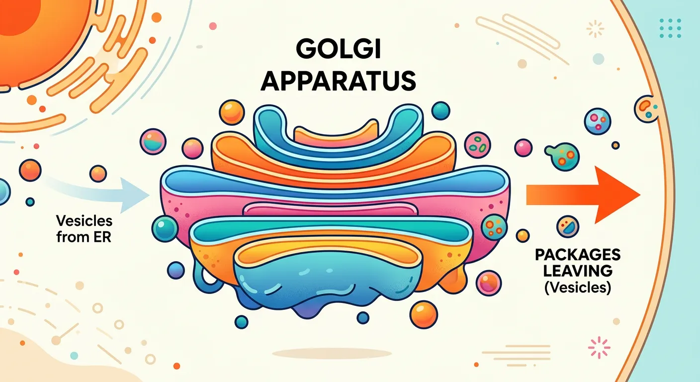
“Think of the Golgi Apparatus as the cell’s Amazon Delivery Center. It takes the raw products from the ER, modifies them, tags them with chemical labels, and ships them to their correct destination. It’s the final post-office ensuring everything reaches the right address inside the cell.”
Camillo Golgi first observed these densely stained reticular structures near the nucleus in 1898. The Golgi apparatus (or Golgi complex) consists of many flat, disc-shaped sacs or cisternae of 0.5μm to 1.0μm diameter. These are stacked parallel to each other. A unique feature of the Golgi is its distinct polarity. It has a convex 'cis' or forming face (oriented toward the nucleus) and a concave 'trans' or maturing face (oriented toward the plasma membrane). Although the cis and trans faces are different, they are interconnected.
The primary function of the Golgi apparatus is packaging. Materials synthesized by the ER (proteins and lipids) are delivered to the cis face of the Golgi in the form of vesicles. As these materials move through the cisternae toward the trans face, they undergo 'post-translational modification.' The most notable modification is glycosylation—the addition of sugar moieties to proteins and lipids to form glycoproteins and glycolipids, respectively. This is why the Golgi is often called the 'post office' or 'shipping department' of the cell.
Furthermore, the Golgi apparatus is the site where lysosomes are formed. Vesicles containing hydrolytic enzymes bud off from the trans face. Because the Golgi is so intimately involved with the ER and lysosomes, they are all considered part of the 'Endomembrane System.' This coordination ensures that a protein produced in the RER is correctly modified, tagged, and sent to its final destination, whether that is inside the cell or secreted outside.
Quick Revision Points
- Discovery: Camillo Golgi (1898).
- Polarity: Cis face (forming, near ER) vs. Trans face (maturing, near membrane).
- Glycosylation: Major site for the formation of glycoproteins and glycolipids.
- Packaging: Modifies and packages ER products into vesicles for transport.
- Endomembrane System: Works in close coordination with ER and lysosomes.
NEET Exam Angle
- Cis vs Trans: NEET often asks which face receives vesicles from the ER (Answer: Cis).
- Glycoproteins/Glycolipids: This is the most common functional question regarding the Golgi.
- Directionality: Remember the flow: ER → Cis Golgi → Trans Golgi → Target/Secretory vesicle.
| Face of Golgi | Shape | Orientation | Primary Role |
|---|---|---|---|
| Cis Face | Convex | Towards Nucleus/ER | Receiving vesicles |
| Trans Face | Concave | Towards Plasma Membrane | Shipping/Maturing |
| Medial Cisternae | Flat | Between Cis and Trans | Processing/Glycosylation |
06Lysosomes: Hydrolytic Enzymes and Cellular Waste Management
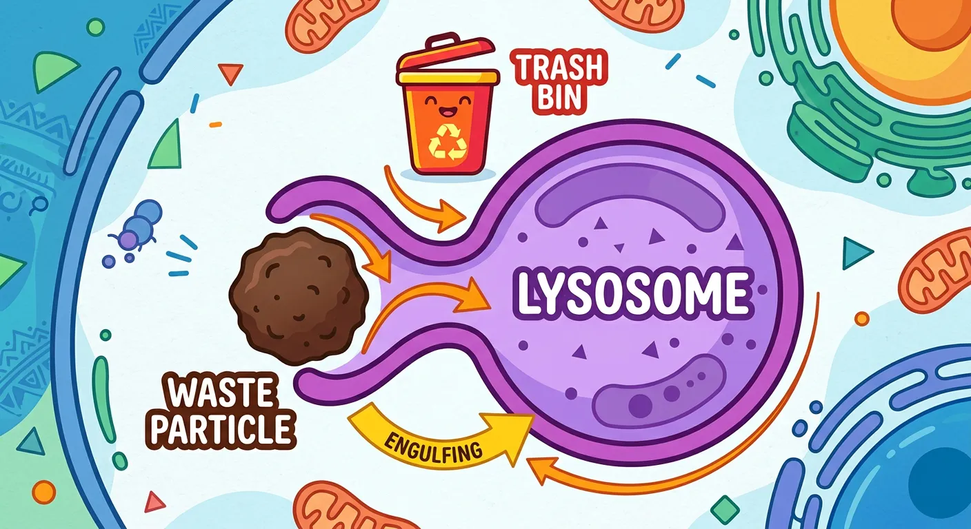
“Lysosomes are the cell’s dedicated Waste Management squad. Filled with powerful digestive enzymes, they break down worn-out parts and foreign invaders. They are essentially 'suicidal bags' that keep the city clean by recycling trash, ensuring the cell remains in peak, healthy condition for your body.”
First discovered by Christian de Duve in 1955, lysosomes are membrane-bound vesicular structures formed by the process of packaging in the Golgi apparatus. They are essentially the 'digestive system' of the cell. These tiny bags are filled with a variety of hydrolytic enzymes (acid hydrolases) including lipases, proteases, glycosidases, and carbohydrases. A key characteristic of these enzymes is that they are optimally active at an acidic pH (around pH 4.5 to 5.0). The lysosomal membrane contains specialized proton pumps that actively transport H+ ions into the lysosome to maintain this internal acidity, protecting the rest of the cytoplasm from accidental digestion.
Lysosomes perform several vital roles. They are involved in 'intracellular digestion,' breaking down macromolecules like proteins, lipids, and nucleic acids into their monomeric subunits. They also engage in 'autophagy' (self-eating), where they digest worn-out organelles such as mitochondria or ER fragments, recycling their components for the cell's use. In certain conditions, if a cell is damaged beyond repair or infected, lysosomes may burst, releasing their enzymes and digesting the entire cell. This programmed cell destruction is why they are famously called 'suicidal bags.'
Furthermore, lysosomes exhibit polymorphism, existing in different states like primary lysosomes (newly formed), secondary lysosomes (fused with food vacuoles), and residual bodies (containing undigested waste). In specialized white blood cells like macrophages, lysosomes fuse with phagosomes to destroy pathogens. This connection to the immune system highlights the lysosome's importance in maintaining overall health, not just cellular hygiene.
Quick Revision Points
- Discovery: Christian de Duve (1955).
- Enzymes: Acid Hydrolases (lipases, proteases, carbohydrases, nucleases).
- Optimal pH: Acidic (approx. 4.5–5.0), maintained by active proton pumps.
- Origin: Formed by budding from the trans-face of the Golgi apparatus.
- Functions: Digestion of macromolecules, autophagy (organelle recycling), and phagocytosis.
- Polymorphism: They exist as primary, secondary, and tertiary (residual) forms.
NEET Exam Angle
- Acidic pH: A very common question focuses on why lysosomal enzymes don't digest the whole cell normally (Answer: they require an acidic pH to function, which is only inside the lysosome).
- Enzyme Types: Match the following questions often pair 'Hydrolases' with 'Lysosomes.'
- RBCs: Remember that mature mammalian erythrocytes lack lysosomes.
- Terminology: Be familiar with 'Autolysis'—the self-destruction of cells by lysosomal enzymes.
07Vacuoles: Osmoregulation and Nutrient Storage in Plant Cells
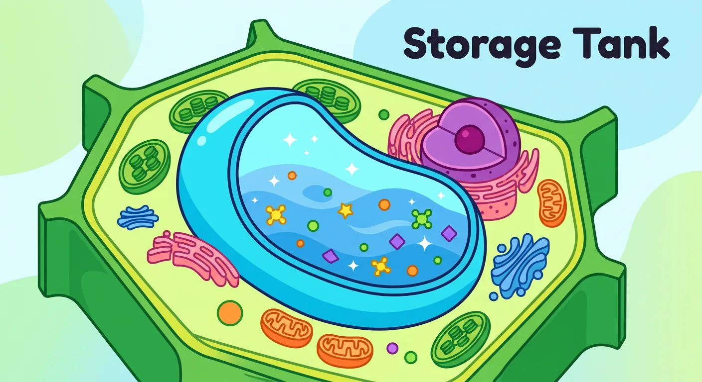
“Lastly, meet the Vacuole. In plant cells, it’s a massive water storage tank that keeps the plant upright and sturdy. It stores water, nutrients, and even waste. It's the ultimate pantry that ensures the cell stays plump, hydrated, and ready for whatever environmental challenge comes its way.”
In the plant world, the vacuole is often the most prominent feature of the cell, sometimes occupying up to 90% of the cell volume. The vacuole is a membrane-bound space found in the cytoplasm. In plants, it is bounded by a single, semi-permeable membrane called the tonoplast. The tonoplast is remarkable because it facilitates the transport of a number of ions and other materials against concentration gradients into the vacuole. This results in the concentration of these materials being significantly higher in the vacuole than in the surrounding cytoplasm.
Vacuoles serve as storage depots for water, sap, excretory products, and other materials not useful for the cell. The high concentration of solutes inside the vacuole draws water in via osmosis, creating turgor pressure. This pressure pushes the plasma membrane against the cell wall, making the cell turgid—this is what keeps herbaceous plants upright. Additionally, many plant vacuoles contain anthocyanin pigments, which give flowers their red, blue, or purple colors.
In other organisms, vacuoles take on different specialized roles. In Amoeba, the 'contractile vacuole' is vital for osmoregulation and excretion, pumping out excess water. In many protists, 'food vacuoles' are formed by engulfing food particles. Despite their simple appearance, vacuoles are dynamic organelles that adapt to the physiological needs of the organism, acting as both a pantry and a pressure-regulating system.
Quick Revision Points
- Tonoplast: The single membrane of the plant vacuole; handles active transport.
- Plant Content: Contains water, sap, excretory products, and pigments.
- Turgidity: Provides structural support in plants via turgor pressure.
- Contractile Vacuole: Found in Amoeba; used for osmoregulation.
- Food Vacuole: Found in protists; used for digestion of engulfed food.
NEET Exam Angle
- Tonoplast Transport: Crucial fact: Ion concentration is higher inside the vacuole than in the cytoplasm due to the tonoplast's active transport.
- Amoeba: Remember that the contractile vacuole is analogous to the human kidney in terms of osmoregulation.
- Percentage: Recall the '90% volume' statistic for mature plant cells—it's a favorite for 'True/False' style questions.
Recommended Reading
Explore related Biology topics to build deeper chapter connections for NEET.
- Cell Theory · Topic 3.1
- Golgi Bodies · Topic 3.10
- Lysosomes · Topic 3.11
- Vacuoles · Topic 3.12
- Plastids · Topic 3.15
- Prokaryotic and Eukaryotic Cell · Topic 3.2
- Jump to Key Terms (Quick Revision)
- Review Common NEET Mistakes
- Read Topic FAQs
- Check PYQ Pattern Notes
- Practice NEET MCQs
- Solve NEET PYQs
📚 Key Terms
⚠️ Common NEET Mistakes
- 1Thinking all organelles have the same type of ribosomes; remember mitochondria have 70S while the cytoplasm has 80S.
- 2Confusing the direction of transport in the Golgi; vesicles always enter at the 'cis' face and exit from the 'trans' face.
- 3Assuming all plant cells have one giant vacuole; while common, some young or specialized plant cells may have several smaller ones.
- 4Forgetting that the cell membrane contains carbohydrates (glycoproteins/glycolipids), not just lipids and proteins.
- 5Believing mitochondria produce 'energy' from nothing; they convert chemical energy from glucose into a usable form (ATP).
📝 NEET PYQ Pattern
In NEET 2018–2024, questions frequently focus on matching organelles to their specific functions (e.g., Golgi-glycosylation, SER-lipid synthesis). There is also a consistent trend of asking about the structural components of the endomembrane system and the semi-autonomous nature of mitochondria.
❓ Frequently Asked Questions
Why are Mitochondria and Chloroplasts called semi-autonomous organelles?
They are called semi-autonomous because they possess their own genetic material (circular DNA) and protein-synthesizing machinery (70S ribosomes). This allows them to produce some of their own proteins and replicate independently by fission, although they still depend on the nuclear genome for many other proteins and functions.
What is the difference between the 'cis' and 'trans' faces of the Golgi apparatus?
The 'cis' face is the convex, forming face oriented toward the nucleus/ER, which receives transport vesicles. The 'trans' face is the concave, maturing face oriented toward the plasma membrane, where modified materials are packaged into vesicles and shipped out.
How does the Rough ER differ from the Smooth ER in both structure and function?
Rough ER (RER) has ribosomes on its surface and consists mainly of flattened cisternae; its primary function is protein synthesis and secretion. Smooth ER (SER) lacks ribosomes and consists of tubules; its primary function is the synthesis of lipids, steroid hormones, and detoxification of drugs.
Why are lysosomes known as the 'suicidal bags' of the cell?
Lysosomes contain powerful hydrolytic enzymes that can digest all organic materials. If a cell is severely damaged or reaches the end of its life cycle, the lysosome may burst, releasing these enzymes into the cytoplasm and causing the cell to digest itself (autolysis).
What is the role of the Tonoplast in plant cell vacuoles?
The tonoplast is the single membrane surrounding the plant vacuole. Its primary role is to actively transport ions and other materials against their concentration gradient from the cytoplasm into the vacuole, maintaining high osmotic pressure and cell turgidity.
Which cell organelles are considered part of the endomembrane system?
The endomembrane system includes the Endoplasmic Reticulum (ER), Golgi complex, Lysosomes, and Vacuoles. These organelles are grouped together because their functions are highly coordinated and they share membrane components through vesicle transport.
Written By
NEET Content Strategist & Biology Expert
Sangita Kumari is a NEET educator and content strategist with over 6 years of experience teaching Biology, Chemistry, and Physics to Class 11 and 12 aspirants. She helps bridge the gap between traditional NCERT preparation and modern AI-powered learning. Her content is trusted by thousands of NEET aspirants across India.