🎬 Video Lesson Available
Watch the full 7-slide video lesson for Animal Tissues with AI teacher narration and visual explanations.
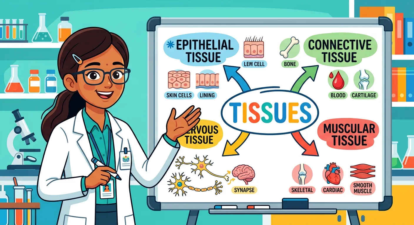
01Foundations of Structural Organisation: The Four Pillars of Animal Tissues
“Welcome, future doctors! Think of your body like a grand skyscraper. Animal tissues are the four types of construction materials that make it all work. We have the outer skin bricks, the supporting cement, the moving wires, and the control cables. Let us break them down!”
In the vast world of multicellular organisms, life isn't just about a collection of cells; it is about how those cells cooperate. Think of a single-cell Amoeba—it performs every task, from digestion to excretion, within one boundary. However, as animals became more complex through evolution, a 'division of labour' became necessary. This led to the formation of tissues: groups of similar cells, along with intercellular substances, performing a specific function. Understanding animal tissues is like learning the architectural blueprints of life. It connects your knowledge of basic cell biology to the complex human physiology you will study in later chapters. The term 'histology' specifically refers to the microscopic study of these tissues, a field that was revolutionized by the development of high-resolution microscopy.
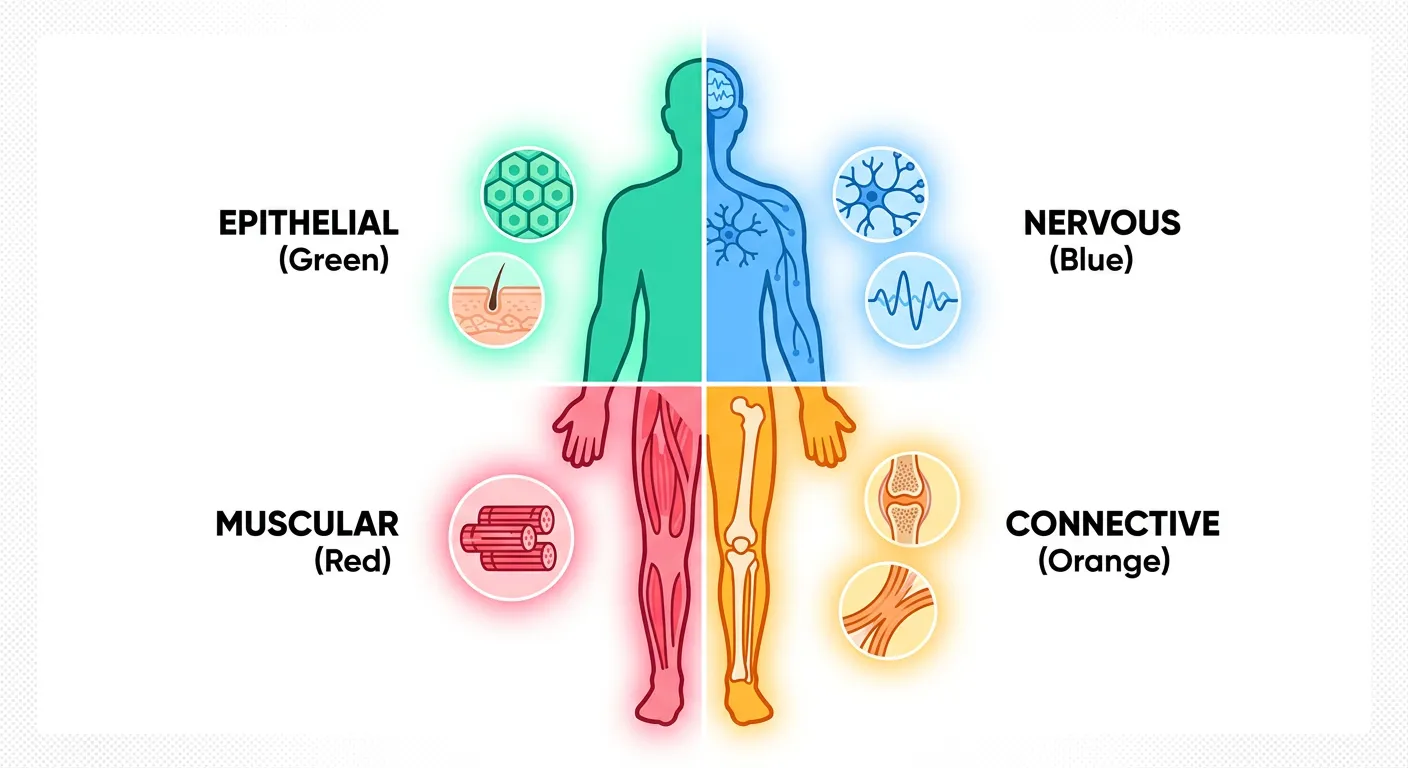
To grasp this hierarchy-and-systematics), imagine building a skyscraper. You don't just throw bricks together; you need different materials for different purposes. In the animal kingdom, these materials are classified into four fundamental types: Epithelial, Connective, Muscular, and Nervous. Each tissue type evolved from specific embryonic germ layers—Ectoderm, Mesoderm, or Endoderm—to meet a specific physiological demand. Whether it is the protective barrier of your skin or the rapid electrical signals of your brain, every function is rooted in tissue specialization. This organization allows complex animals to maintain homeostasis, a state of internal balance, and survive in diverse environments. This concept of structural organization is a recurring theme in NEET, where the focus is on how form dictates function.
Quick Revision Points
- Tissues Definition: Groups of similar cells plus intercellular matrix working toward a common goal.
- Division of Labour: Evolution of tissues allowed for greater body size and functional complexity in animals.
- The Four Pillars: Epithelial (covering), Connective (support), Muscular (movement), and Nervous (control).
- Hierarchy: Tissue systems bridge the gap between individual cell organelles and full-fledged organ systems.
NEET Exam Angle
- Focus on Definition: NEET often tests the concept that tissues include both cells and 'intercellular substances'.
- Germ Layer Origins: Note that Epithelial tissue can arise from all three germ layers, whereas others are more specific.
- Interconnectivity: Remember that Unit 2 (Structural Organisation) provides the morphological basis for Unit 5 (Human Physiology).
| Tissue Type | Primary Function | Basic Example | Embryonic Origin |
|---|---|---|---|
| Epithelial | Protection, Secretion | Skin, Gut lining | Ecto/Meso/Endoderm |
| Connective | Binding, Support | Bone, Blood, Tendons | Mesoderm |
| Muscular | Locomotion | Biceps, Heart wall | Mesoderm |
| Nervous | Communication | Brain, Spinal cord | Ectoderm |
02Epithelial Tissue: The Protective Lining and Cellular Packaging
“First, the Epithelial tissue—your body’s protective packaging! Like floor tiles in your home, they cover surfaces and line cavities. From flat squamous scales to tall columnar pillars, these cells are our first line of defense. They protect, absorb, and filter everything that enters your system.”
Epithelial tissue, or epithelium, serves as the body's versatile wrapper. It has a free surface, which faces either a body fluid or the outside environment, providing a compact covering or lining for various body parts. One of the most distinctive features of epithelial tissue is that the cells are compactly packed with very little intercellular matrix. This 'tile-like' arrangement makes it an excellent barrier. It rests upon a non-cellular basement membrane, composed of glycoproteins and collagen, which provides structural support and serves as a selective filter for nutrients since epithelial tissues themselves are avascular (lacking blood vessels). They rely on the underlying connective tissue for their metabolic needs.
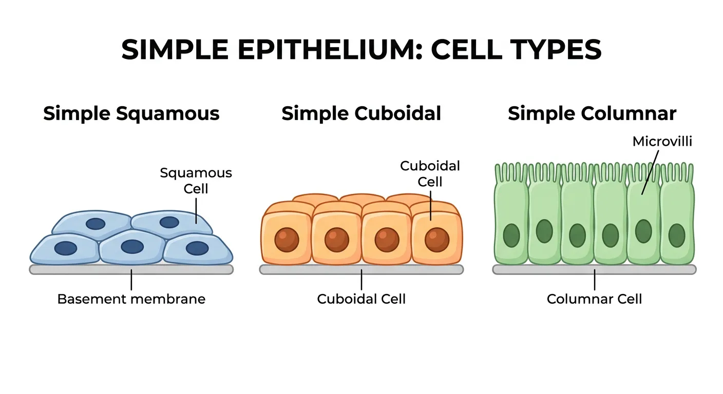
We classify epithelia into two main categories: Simple and Compound. Simple epithelium consists of a single layer of cells and is usually found where absorption or filtration is required, such as in the linings of body cavities, ducts, and tubes. Compound (or stratified) epithelium consists of two or more cell layers and primarily functions in protection against chemical and mechanical stresses—think of your skin or the lining of the buccal cavity! Furthermore, we distinguish simple epithelium based on cell shape: squamous (flat), cuboidal (cube-like), and columnar (tall and slender). These shapes aren't just for show; they dictate how the organ functions. For instance, the thinness of squamous cells in the lung alveoli facilitates the rapid diffusion of gases, whereas the microvilli-covered columnar cells in the intestine maximize nutrient absorption.
Quick Revision Points
- Squamous Epithelium: Flat cells with irregular boundaries; found in blood vessel walls and air sacs of lungs.
- Cuboidal Epithelium: Cube-shaped cells; commonly found in ducts of glands and tubular parts of nephrons.
- Columnar Epithelium: Tall cells with nuclei at the base; line the stomach and intestine for secretion and absorption.
- Cell Junctions: Tight junctions prevent leakage; Adhering junctions cement cells; Gap junctions allow communication.
NEET Exam Angle
- Ciliated Epithelium: High-yield topic. Remember their location in bronchioles and fallopian tubes to move particles or eggs.
- Glandular Epithelium: Distinguish between Unicellular (Goblet cells) and Multicellular (Salivary glands).
- Basement Membrane: Understand that it is non-cellular and serves as the anchor for all epithelial layers.
| Epithelium Type | Location | Key Function | Modification |
|---|---|---|---|
| Simple Squamous | Alveoli, Blood vessels | Diffusion boundary | Thin, flat cells |
| Simple Cuboidal | Kidney tubules (PCT) | Secretion & Absorption | Brush border (Microvilli) |
| Simple Columnar | Stomach lining | Absorption | Tall, slender cells |
| Ciliated | Fallopian tubes | Particle movement | Hair-like Cilia |
03Connective Tissues: The Biological Glue and Structural Framework
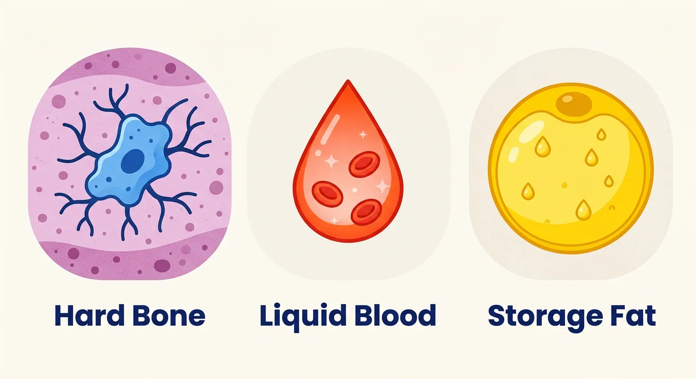
“Next, the glue that holds everything together: Connective tissue. It is the most abundant type! Whether it is rigid bone, flowing blood, or soft storage fat, this tissue provides structural support and transports nutrients. It is the connective bridge between all your vital organs.”
Connective tissue is the most abundant and widely distributed tissue in the body of complex animals. Its primary role is exactly what the name suggests: linking and supporting other tissues and organs. Unlike epithelial tissue, where cells are packed tight, connective tissue is defined by its vast extracellular matrix. This matrix consists of ground substance (often modified polysaccharides like hyaluronic acid) and protein fibers like collagen and elastin. Collagen provides immense tensile strength, while elastin allows the tissue to stretch and recoil, ensuring our organs stay in place yet remain flexible during movement.
We categorize connective tissues into three main types: Loose, Dense, and Specialized. Loose connective tissue, like areolar and adipose tissue, has cells and fibers loosely arranged in a semi-fluid ground substance. Areolar tissue often serves as the support framework for epithelium. Adipose tissue is a specialized reservoir for fats, located mainly under the skin. Dense connective tissues, such as tendons (attaching muscle to bone) and ligaments (attaching bone to bone), contain fibers packed tightly to resist tension. Dense regular tissue has fibers in parallel rows, while dense irregular tissue (found in the skin) has a mesh-like arrangement. Finally, specialized connective tissues include cartilage, bone, and blood. Blood is unique because it is a fluid connective tissue that lacks structural fibers but contains plasma, RBCs, WBCs, and platelets, serving as the body's primary highway for transport.
Quick Revision Points
- Fibroblasts: The main cells that secrete fibers (collagen/elastin) in most connective tissues.
- Adipose Tissue: Located mainly beneath the skin; stores nutrients as fats and provides thermal insulation.
- Cartilage vs. Bone: Cartilage is solid and pliable (chondrocytes), while bone is hard and non-pliable (osteocytes) due to calcium salts.
- Blood: The only connective tissue without structural fibers in its normal physiological state.
NEET Exam Angle
- Tendons vs. Ligaments: This is a classic MCQ trap. Remember: Tendons = Muscle to Bone (TMB); Ligaments = Bone to Bone (LBB).
- Matrix Composition: Pay attention to the types of fibers present (e.g., White fibers are collagen, Yellow are elastin).
- Dense Irregular: Remember its presence in the dermis of the skin, providing multi-directional strength.
| Connective Type | Key Cells | Distinctive Feature | Matrix State |
|---|---|---|---|
| Areolar | Fibroblasts, Macrophages | Support framework | Semi-fluid |
| Adipose | Adipocytes | Fat storage area | Semi-fluid |
| Dense Regular | Collagen fibers in rows | Tendons/Ligaments | Fibrous |
| Specialized | Osteocytes (Bone) | Calcified matrix | Hard/Solid |
04Muscular Tissue: Contractility and the Mechanics of Movement
“Ready to move? Meet Muscular tissue! These are your body’s engines. Skeletal muscles help you sprint, smooth muscles power your digestion, and the special cardiac muscle keeps your heart beating non-stop. They contract and relax to turn chemical energy into the physical power of your body.”
Muscular tissue is specialized for one thing: contraction. Each muscle is made of many long, cylindrical fibers arranged in parallel arrays. These fibers are composed of fine fibrils called myofibrils, which contain the contractile proteins actin and myosin. When these fibers contract (shorten) and then relax (lengthen), they bring about the movement of body parts or the internal organs. This process is highly energy-intensive, requiring the conversion of chemical energy from ATP into mechanical work. Because of this high metabolic rate, muscle tissues are richly supplied with blood vessels and mitochondria.
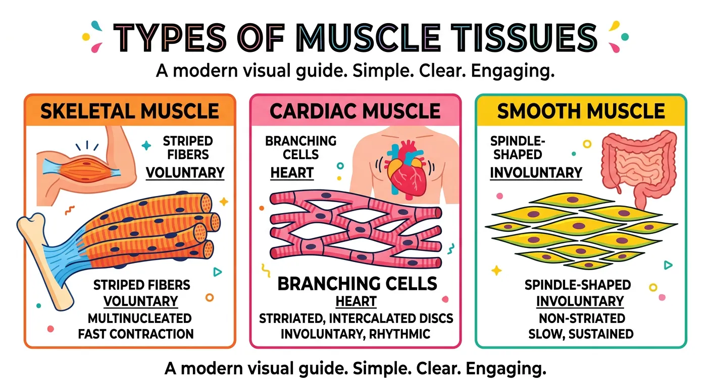
There are three distinct types of muscle tissue: Skeletal, Smooth, and Cardiac. Skeletal muscle is closely attached to skeletal bones and is 'striated' (striped) in appearance due to the organized arrangement of myofibrils. It is under voluntary control, meaning you decide when to move your limb. These cells are often multinucleated (syncytium). Smooth muscle, found in the walls of internal organs like the stomach and blood vessels, is 'non-striated' and involuntary. The cells are fusiform, meaning they taper at both ends. You cannot consciously speed up your digestion or change your blood pressure! Lastly, Cardiac muscle is unique to the heart. It features branching patterns and specialized junctions called intercalated discs. These discs act as 'boosters' that allow electrical signals to spread rapidly so that the heart contracts as a single, coordinated unit—a vital feature for maintaining rhythmic blood circulation.
Quick Revision Points
- Striations: Present in Skeletal and Cardiac muscles; absent in Smooth muscles.
- Voluntary Control: Only Skeletal muscle is voluntary.
- Intercalated Discs: Unique to Cardiac muscle; function as communication boosters for synchronous contraction.
- Fusiform Shape: Smooth muscle cells are spindle-shaped (tapering at both ends) with a single central nucleus.
NEET Exam Angle
- Cardiac Muscle Syncytium: NEET loves asking about the 'syncytium-like' behavior of cardiac cells due to gap junctions.
- Location Identification: Be ready to identify which muscle type is found in specific organs (e.g., iris of the eye or wall of the aorta).
- Myofibrils: Understand that the structural unit of contraction is the sarcomere (covered in the Locomotion chapter).
| Muscle Type | Appearance | Control | Location | Cell Nuclei |
|---|---|---|---|---|
| Skeletal | Striated | Voluntary | Attached to bones | Multinucleated |
| Smooth | Spindle-shaped | Involuntary | Visceral organs | Uninucleated |
| Cardiac | Striated/Branched | Involuntary | Heart Wall | Uninucleated |
| Myocyte | Contractile unit | Variable | Throughout body | N/A |
05Nervous Tissue: Neural Communication and The Command Center
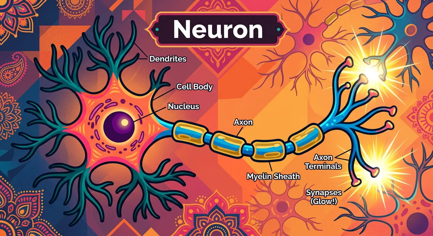
“Now, for the body's high-speed internet: Nervous tissue. The star here is the neuron. It carries electrical impulses faster than a text message! It senses the world, processes information, and tells your muscles when to move. It is the command center of your entire existence.”
Nervous tissue exerts the greatest control over the body's responsiveness to changing conditions. While the other tissues provide structure and movement, nervous tissue provides intelligence, coordination, and rapid communication. The fundamental unit of this tissue is the neuron. Neurons are 'excitable' cells, meaning they can generate and conduct electrical impulses known as action potentials. A typical neuron consists of a cell body (cyton) containing the nucleus, dendrites (short, branched fibers which receive incoming signals), and a long axon (a single fiber which transmits signals away from the cell body). This specialized structure is perfectly adapted for rapid, long-distance communication within the complex animal body.
However, neurons aren't alone in this tissue. Nervous tissue also contains a massive number of neuroglial cells. Interestingly, neuroglia make up more than one-half the volume of neural tissue in our body! While they don't conduct impulses themselves, they are absolutely crucial for protecting and supporting neurons. They act as the 'pit crew' for the neurons, providing electrical insulation (like the myelin sheath produced by Schwann cells), metabolic support, and maintaining the chemical environment (homeostasis) of the brain and spinal cord. When a neuron is stimulated, an electrical disturbance is generated which travels along its plasma membrane to the axon terminals. This leads to the release of neurotransmitters at the synapse, allowing the message to jump to the next neuron or to a muscle cell, triggering a physiological response. This intricate network forms the basis of our sensory perception, memory, and voluntary actions.
Quick Revision Points
- Neuron: The structural and functional unit of the nervous system, capable of excitability and conductivity.
- Neuroglia: Support cells (like Astrocytes and Microglia) that do not conduct impulses but protect and nourish neurons.
- Impulse Transmission: A process where electrical signals move from the dendrite through the cyton to the axon.
- Volume Ratio: Neuroglial cells occupy more than 50% of the total neural tissue volume in humans.
NEET Exam Angle
- Neuroglia Importance: Remember that neuroglia are the most numerous cells in the brain—a common fact-based MCQ in NEET.
- Impulse Path: Understand the unidirectional flow: Dendrite -> Cell Body -> Axon -> Synapse -> Target cell.
- Regeneration: Note that mature neurons have very limited capacity for cell division, which is why nerve damage is often permanent.
| Component | Function | Conductivity | Relative Abundance |
|---|---|---|---|
| Neuron | Transmits electrical impulses | Yes | High functionality |
| Neuroglia | Support, protection, insulation | No | >50% by volume |
| Dendrite | Receives incoming stimuli | Yes | Multiple per neuron |
| Axon | Carries impulse away | Yes | Usually one per neuron |
06Organ and Organ Systems: Tissue Synergy in Biological Functions
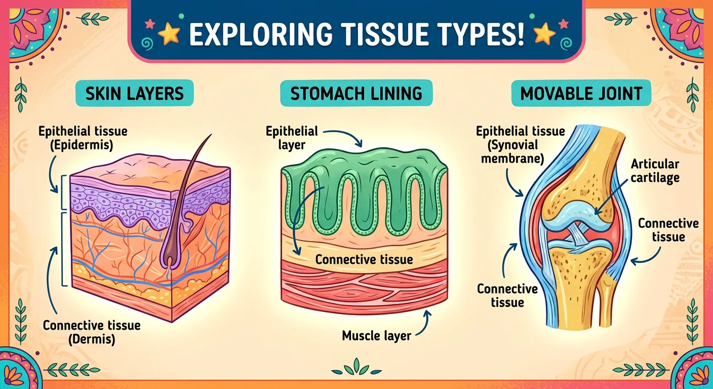
“Tissues rarely work alone. Just like a perfectly cooked Biryani needs rice, spices, and meat together, your organs like the stomach are made of multiple tissues. The epithelial lining digests, the muscle churns, and connective tissue holds the structural shape. Teamwork makes the dream work!”
Individual tissues are remarkable, but their true power is realized when they combine to form organs. An organ is a group of different tissues working together in a coordinated manner to perform a specific, complex function. Take the human stomach, for example. It isn't just one type of tissue; it's a sophisticated biological machine. Its inner lining is epithelial (specifically glandular columnar epithelium for the secretion of digestive juices), its walls contain layers of smooth muscle (to churn food), it is wrapped in connective tissue (providing structural integrity and a pathway for blood vessels), and it is permeated by nervous tissue (to signal hunger and regulate the rhythm of digestion). This integration of all four tissue types is the hallmark of higher biological organization.
This organization into organs and eventually organ systems (like the digestive, circulatory, or respiratory systems) allows for a massive 'division of labour'. This efficiency is essential for the survival of complex multicellular animals, as it allows specialized cells to focus on specific tasks. For instance, the heart is a muscular pump, but its histology reveals cardiac muscle for contraction, connective tissue for the valves and structural scaffolding, and epithelial tissue for the smooth inner lining (endocardium). When you study the anatomy of any organ, you are essentially studying histology—the study of tissues—on a macro scale. This interdependency ensures that the internal environment remains stable despite changes in the external world. Mastering the transition from tissue to organ is key to understanding the 'Structural Organisation' unit as a whole, as it sets the stage for every physiological process you will encounter in your medical studies.
Quick Revision Points
- Organ Definition: Multiple tissue types (Epithelial, Connective, Muscular, Nervous) working together.
- Division of Labour: Specialization at the organ level increases overall biological efficiency and survival rates.
- Morphology vs. Anatomy: Morphology is the study of external forms; Anatomy is the study of internal tissue and organ organization.
- Example Integration: The heart and stomach utilize all four basic tissue types to function effectively.
NEET Exam Angle
- Stomach Histology: Remember the four layers: Serosa (Connective), Muscularis (Muscle), Submucosa, and Mucosa (Epithelial).
- Complexity: NEET often asks about the highest level of organization—the 'Organ System' level vs the 'Tissue' level.
- Tissue Synergy: Understand how nervous tissue (vagus nerve) triggers muscular contraction in the gut walls.
| Organ | Primary Tissue Layer | Secondary Tissue Layer | Functional Outcome |
|---|---|---|---|
| Stomach | Mucosa (Epithelial) | Muscularis (Smooth Muscle) | Digestion/Churning |
| Heart | Myocardium (Cardiac Muscle) | Endocardium (Epithelial) | Blood Pumping |
| Skin | Epidermis (Epithelial) | Dermis (Connective/Nervous) | Protection/Sensation |
| Kidney | Nephrons (Cuboidal Epithel.) | Connective Stroma | Filtration/Excretion |
07NEET Preparation Strategy: Master Animal Histology through Conceptual Clarity
“You have mastered the basics of Animal Tissues! Remember, biology is not about memorizing; it is about seeing how your own body functions. Keep exploring, keep questioning, and keep prepping for that NEET success. You have got this, champions! See you in the next lesson.”
To excel in the NEET Biology section, 'Animal Tissues' should be approached through visualization and comparison. This chapter is a critical bridge; it connects the 'Cell' unit to the 'Human Physiology' unit. The most effective way to study is to use charts and diagrams to map locations. Most NEET questions on this topic are direct, but they often test your precision—your ability to match a specific tissue type with its exact location in the body. For instance, knowing that ciliated epithelium is in the fallopian tubes is a classic high-yield fact that appears frequently in matching-type questions. You must move beyond rote memorization and understand the logic: why is a specific tissue located there? (e.g., Squamous for thin diffusion in the lungs).
Use memory hooks and mnemonics to keep the details straight. For example, think of 'Tight Junctions' as 'Leaky-proof seals' and 'Gap Junctions' as 'Communication Tunnels'. When studying connective tissue, always visualize the matrix—is it fluid like blood, semi-solid like cartilage, or hard like bone? Practice identifying the diagrams directly from the NCERT textbook, as these illustrations are frequently reproduced exactly in the exam papers. Pay close attention to the labels of the fibroblasts, mast cells, and collagen fibers in the areolar tissue diagram. Finally, remember that consistency is key. Reviewing these tissue types and their specific locations regularly will make the complex physiology of the heart, lungs, and kidneys much easier to understand later on. You have the tools and the intellectual capacity; now apply them with the precision of a surgeon! Focus on previous year questions (PYQs) to understand the pattern of 'Assertion and Reason' questions that are becoming more common in this specific chapter.
Quick Revision Points
- Focus on NCERT Diagrams: They are the primary source for visual MCQs and labeling questions.
- Location Charts: Create a master table of all epithelial types and their specific anatomical locations.
- Junction Types: Understand the functional difference between Tight (barrier), Adhering (cement), and Gap (channel) junctions.
- High-Yield Terms: Fibroblasts (fiber-secreting), Chondrocytes (cartilage), Osteocytes (bone), and Intercalated discs (cardiac).
NEET Exam Angle
- Trend Analysis: Questions often repeat on Ciliated epithelium locations and the components of the Connective tissue matrix.
- Conceptual Clarity: Don't just memorize; understand 'form follows function' (e.g., columnar cells have nuclei at the base to allow space for secretions).
- Matching Type MCQs: Practice matching columns of tissue types to functions or locations to improve speed and accuracy.
| Junction Type | Logic Hook | Primary Function | Significance |
|---|---|---|---|
| Tight | 'Stop Leak' | Stops substances leaking across | Prevents gut leaks |
| Adhering | 'Cell Glue' | Cements neighboring cells | Mechanical stability |
| Gap | 'Channel' | Rapid transfer of ions | Syncytial contraction |
Recommended Reading
Explore related Biology topics to build deeper chapter connections for NEET.
- Morphology and Modifications · Topic 2.1
- Families · Topic 2.10
- Frog Morphology · Topic 2.12
- Digestive System · Topic 2.13
- Circulatory System · Topic 2.14
- Respiratory System · Topic 2.15
- Jump to Key Terms (Quick Revision)
- Review Common NEET Mistakes
- Read Topic FAQs
- Check PYQ Pattern Notes
- Practice NEET MCQs
- Solve NEET PYQs
📚 Key Terms
⚠️ Common NEET Mistakes
- 1Confusing the location of Ciliated Epithelium (Bronchioles) with Squamous Epithelium (Alveoli).
- 2Swapping the functions of Tendons (Muscle-to-Bone) and Ligaments (Bone-to-Bone).
- 3Assuming all connective tissues contain fibers (remember, Blood does not have structural fibers).
- 4Thinking that cardiac muscle is voluntary because it is striated; it is strictly involuntary.
- 5Ignoring the fact that neuroglia make up more than 50% of brain volume, often focusing only on neurons.
📝 NEET PYQ Pattern
In NEET 2018–2024, questions frequently focus on matching tissue types to their specific locations (e.g., bronchioles, fallopian tubes). There is a consistent pattern of asking about cell junctions (Tight vs. Gap) and the structural features of specialized connective tissues like cartilage and bone.
❓ Frequently Asked Questions
What is the main difference between simple and compound epithelium in terms of function?
Simple epithelium consists of a single layer of cells and is specialized for absorption, secretion, and filtration. Compound epithelium has multiple layers and primarily provides protection against mechanical or chemical stress, such as in the skin or oral cavity.
Where exactly is ciliated epithelium located in the human body, and why?
Ciliated epithelium is found in the inner surface of hollow organs like the bronchioles and fallopian tubes. The cilia move in a coordinated fashion to push mucus or particles (in the respiratory tract) or the ovum (in the fallopian tubes) in a specific direction.
How do gap junctions facilitate communication between cells in cardiac muscle?
Gap junctions are specialized channels that connect the cytoplasm of adjacent cells. In cardiac muscle, they allow for the rapid transfer of ions and small molecules, enabling electrical impulses to spread instantly so the heart can contract as a single unit.
Why is blood classified as a connective tissue even though it is fluid?
Blood is classified as a connective tissue because it shares an embryonic origin (mesoderm) with other connective tissues and functions to link and support various organs by transporting nutrients, gases, and hormones, despite lacking the structural fibers found in bone or cartilage.
Which type of animal tissue is most abundant and widely distributed in the body?
Connective tissue is the most abundant and widely distributed tissue in the body, ranging from loose tissues like areolar tissue to specialized forms like bone and blood.
What is the specific role of neuroglial cells compared to neurons in nervous tissue?
Neurons are the excitable cells that generate and conduct electrical impulses for communication. Neuroglial cells are non-excitable support cells that protect, insulate, and nourish neurons, making up more than half the volume of the nervous system.
Written By
NEET Content Strategist & Biology Expert
Sangita Kumari is a NEET educator and content strategist with over 6 years of experience teaching Biology, Chemistry, and Physics to Class 11 and 12 aspirants. She helps bridge the gap between traditional NCERT preparation and modern AI-powered learning. Her content is trusted by thousands of NEET aspirants across India.