🎬 Video Lesson Available
Watch the full 7-slide video lesson for Digestive System with AI teacher narration and visual explanations.
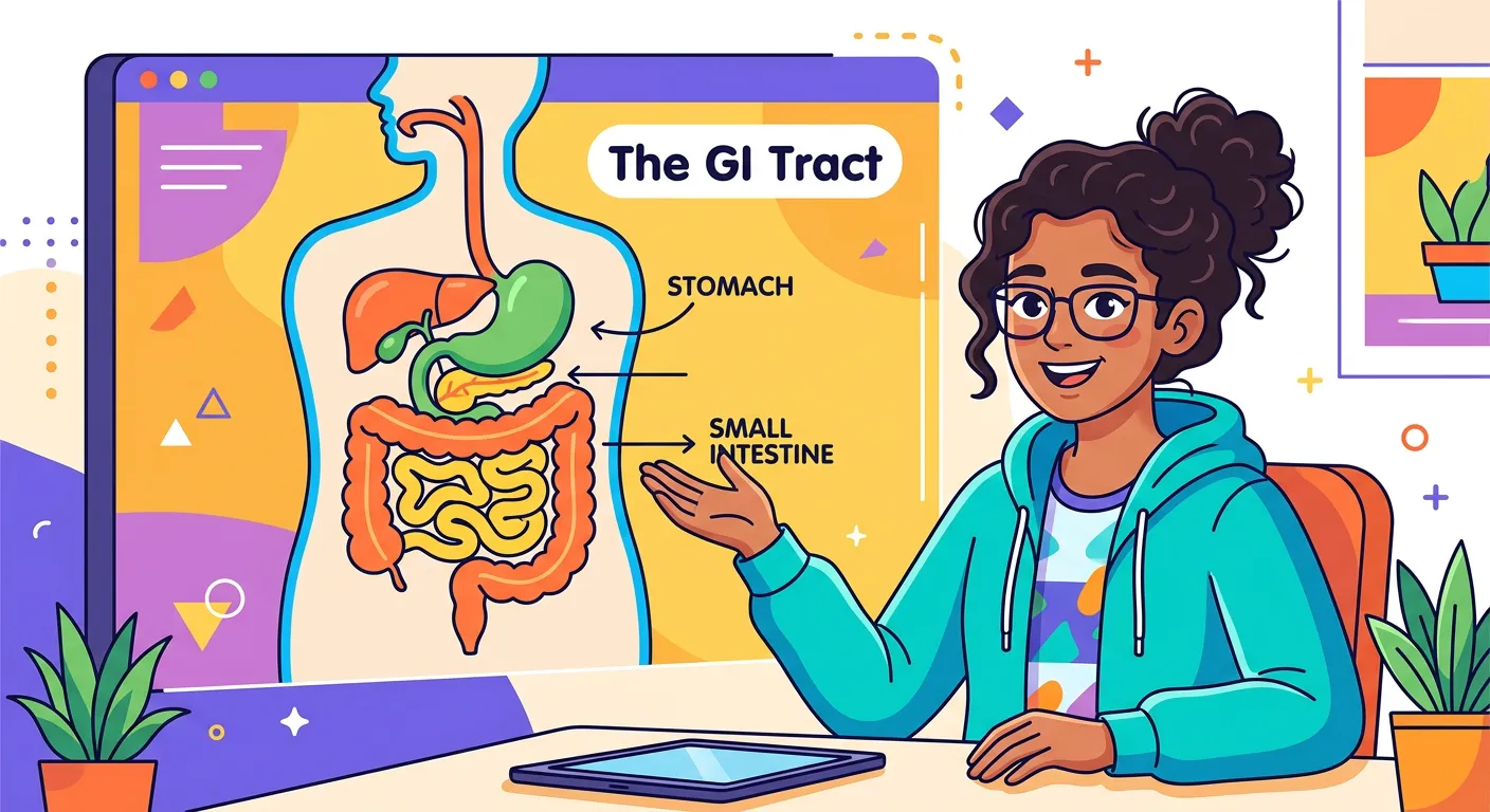
01The Human Digestive System: An Overview of the Biological Assembly Line
“Welcome, future doctors! Think of your digestive system as a sophisticated internal factory, constantly processing the delicious food you eat into pure fuel for your cells. Today, we will journey from the mouth to the finish line, breaking down every step of this incredible biological assembly line.”
Welcome, future doctors! Think of your digestive system as a sophisticated internal factory, constantly processing the food you eat into pure fuel for your cells. Digestion is a complex combination of mechanical and biochemical processes designed to break down large, complex macromolecules into simpler, absorbable units. Without this conversion, your cells would starve even if your stomach were full. The system is essentially an elongated tube, known as the alimentary canal, supplemented by various accessory glands that secrete essential juices to facilitate this transformation. The process begins with ingestion and ends with egestion, following a strictly regulated path that ensures maximum efficiency.
To understand the digestive system, we must first look at the histology of the alimentary canal. From the esophagus to the rectum, the wall of the canal possesses four distinct layers: the serosa (outermost thin mesothelium), the muscularis (smooth muscles arranged into inner circular and outer longitudinal layers), the sub-mucosa (containing nerves, blood, and lymph vessels), and the mucosa (the innermost lining). In the stomach, the mucosa forms irregular folds called rugae, while in the small intestine, it forms villi. This structural hierarchy-and-systematics) is essential for the movement of food via peristalsis and the secretion of digestive enzymes.
The GI tract begins at the mouth and ends at the anus, traversing a unique journey of structural and functional modifications. As we move from the buccal cavity through the esophagus, stomach, small intestine, and finally the large intestine, each section is optimized for specific tasks. While the stomach serves as an acidic holding chamber, the small intestine acts as the primary site for nutrient absorption. Understanding this roadmap is fundamental to mastering human physiology, as it links directly to your understanding of biomolecules—specifically how proteins, carbohydrates, and lipids are dismantled at the molecular level into amino acids, monosaccharides, and fatty acids.
Quick Revision Points
- Digestion involves both mechanical (physical breakdown) and biochemical (enzymatic) processes.
- The wall of the alimentary canal consists of four layers: Serosa, Muscularis, Sub-mucosa, and Mucosa.
- Accessory organs include salivary glands, the liver, and the pancreas.
- The primary goal is converting complex polymers into monomers (e.g., proteins to amino acids).
NEET Exam Angle
- Focus on the sequence of the GI tract organs: Mouth → Pharynx → Esophagus → Stomach → Small Intestine → Large Intestine.
- Understand the histological layers, particularly the location of Brunner's glands in the sub-mucosa of the duodenum.
| Layer | Composition | Key Feature |
|---|---|---|
| Serosa | Mesothelium | Outermost protective layer |
| Muscularis | Smooth Muscle | Responsible for peristalsis |
| Mucosa | Epithelium | Forms villi/rugae for absorption |
02The Buccal Cavity: Mechanical Mastication and Salivary Amylase Action
“Digestion starts before you even swallow! Your teeth perform mechanical breakdown, while salivary glands secrete amylase, an enzyme that turns your complex starch into simpler sugars. That’s why a plain roti starts tasting sweet if you chew it long enough. It is pure chemistry in your mouth!”
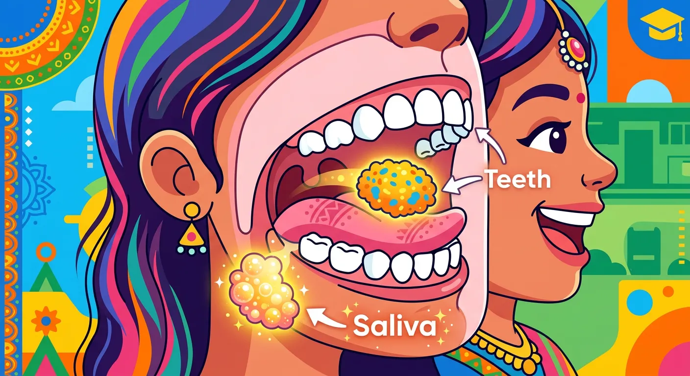
Digestion begins the moment food enters the mouth. Your teeth perform the vital task of mastication—breaking food into smaller pieces. Humans are classified as thecodont (embedded in jaw sockets), diphyodont (two sets of teeth: milk and permanent), and heterodont (different types: incisors, canines, premolars, and molars). The adult human dental formula is 2123/2123, representing two incisors, one canine, two premolars, and three molars in each half of the upper and lower jaws. This structural design maximizes mechanical breakdown efficiency, increasing the surface area for enzymes to act upon. The hard chewing surface of teeth, made of enamel, is the hardest substance in the human body.
Beyond teeth, the tongue and salivary glands play critical roles. The tongue is a freely movable muscular organ attached to the floor of the oral cavity by the frenulum. Its upper surface has small projections called papillae, some of which bear taste buds. Saliva is produced by three pairs of salivary glands: the parotids (cheek), the sub-maxillary/sub-mandibular (lower jaw), and the sub-linguals (below the tongue). Saliva contains electrolytes (Na+, K+, Cl-, HCO3-), mucus, and enzymes like salivary amylase and lysozyme.
Salivary amylase specifically targets starch, initiating chemical digestion by converting approximately 30% of starch into the disaccharide maltose at an optimum pH of 6.8. The result of this process is a lubricated mass called a 'bolus,' which is pushed toward the pharynx. It is fascinating to note that this phase is under neural control; the mere thought, smell, or presence of food triggers the salivary glands. Lysozyme present in saliva acts as an antibacterial agent that prevents infections in the oral cavity, showcasing the mouth's role in both digestion and immunity.
Quick Revision Points
- Teeth types: Thecodont, Diphyodont, and Heterodont (Incisors, Canines, Premolars, Molars).
- Adult dental formula: 2123/2123.
- Salivary amylase acts at pH 6.8 to convert starch to maltose.
- Lysozyme in saliva acts as an antibacterial agent to protect the oral cavity.
NEET Exam Angle
- Expect questions on dental formulas and the specific chemical action of salivary amylase.
- Remember: Digestion of starch begins in the mouth, but protein and lipid digestion do not.
| Component | Function |
|---|---|
| Teeth (Enamel) | Mechanical breakdown and mastication |
| Salivary Amylase | 30% Starch hydrolysis to Maltose |
| Lysozyme | Antibacterial defense mechanism |
03The Esophagus and Peristalsis: The Physics of Food Transport
“The food travels down the esophagus via peristalsis. Imagine squeezing a toothpaste tube from the bottom up—that wave-like muscular contraction is how your food reaches the stomach. It is an involuntary dance of muscles ensuring your food only moves in one direction: down to the fuel tank.”
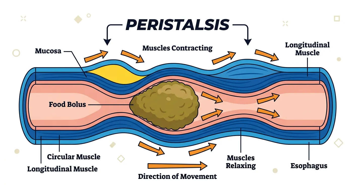
The esophagus is a thin, long tube, approximately 25 cm in length, connecting the pharynx to the stomach. The pharynx serves as a common passage for food and air. During swallowing, a cartilaginous flap called the epiglottis covers the glottis (the opening of the windpipe) to prevent the entry of food into the larynx. Unlike other parts of the GI tract, the esophagus does not secrete digestive enzymes. Its primary job is transport, moving the bolus from the oral cavity to the stomach. This movement occurs through a series of rhythmic, wave-like muscular contractions known as peristalsis.
Peristalsis is facilitated by the coordination of the circular and longitudinal muscles in the muscularis layer. As the bolus enters the esophagus, the muscles behind it contract while the muscles in front relax, ensuring a one-way passage regardless of gravity. This process is involuntary once initiated by the swallowing reflex. Safety and regulation are paramount here. The gastro-esophageal sphincter (a muscular valve) controls the passage of the bolus into the stomach. It acts as a one-way gate that prevents the highly acidic contents of the stomach from splashing back up into the esophagus, a condition commonly known as acid reflux or heartburn.
Mucus secretion from goblet cells is also vital in this region; it acts as a lubricant, reducing friction as the bolus travels down. The histology of the esophagus is unique because its upper third consists of skeletal muscle, the middle third is a mix, and the lower third is entirely smooth muscle. Think of the esophagus as a smooth, high-speed conveyor belt designed specifically for delivery, ensuring the food reaches the 'fuel tank' (the stomach) safely and efficiently without any chemical modification of the nutrients.
Quick Revision Points
- The pharynx is a common passage for food and air; the epiglottis prevents choking.
- Peristalsis involves involuntary wave-like contractions of smooth muscles.
- Mucus provides lubrication to prevent damage to the esophageal lining.
- The gastro-esophageal sphincter prevents the backflow of acidic chyme.
NEET Exam Angle
- Differentiate between the voluntary phase of swallowing and the involuntary phase of peristalsis.
- Note the role of the epiglottis in respiratory-digestive coordination, a common exam topic.
| Feature | Clinical Significance |
|---|---|
| Epiglottis | Prevents food entry into the trachea |
| Gastro-esophageal Sphincter | Prevents GERD (Acid Reflux) |
| Peristalsis | Involuntary propulsion of bolus |
04Gastric Digestion: Hydrochloric Acid and Protein Denaturation
“The stomach is your high-acid chamber. With a pH of around 1.8, the hydrochloric acid kills germs and activates pepsin, the enzyme that shreds proteins. Think of it as a biological blender where food is turned into a creamy, acidic mixture called chyme before heading to the intestines.”
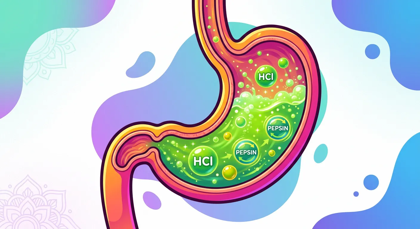
The stomach is a J-shaped muscular bag located in the upper left portion of the abdominal cavity. It is divided into four major regions: the cardiac (where the esophagus enters), fundic, body (main central region), and pyloric (which opens into the first part of the small intestine). The stomach stores food for 4-5 hours. The mucosa of the stomach has gastric glands which contain three main types of cells: mucus neck cells (secrete mucus), Peptic or Chief cells (secrete the proenzyme pepsinogen), and Parietal or Oxyntic cells (secrete HCl and intrinsic factor).
Hydrochloric Acid (HCl) creates a highly acidic environment (pH 1.8), which serves several critical purposes: it kills harmful bacteria entering with food, denatures proteins to expose their peptide bonds, and activates enzymes. Pepsinogen, an inactive proenzyme, is converted into its active form, pepsin, in the presence of HCl. Pepsin is the primary proteolytic enzyme of the stomach, converting proteins into proteoses and peptones (peptides). In infants, the gastric juice also contains rennin, a proteolytic enzyme that helps in the digestion of milk proteins. Small amounts of lipases are also secreted by gastric glands.
While the chemical digestion occurs, the stomach walls perform vigorous churning movements, mixing the food thoroughly with the acidic gastric juice to transform the solid bolus into a semi-digested, creamy mixture called 'chyme.' The mucus and bicarbonates present in the gastric juice play a vital role in lubrication and protection of the mucosal epithelium from excoriation by the highly concentrated hydrochloric acid. Once processing is complete, the pyloric sphincter regulates the release of this chyme into the duodenum in small, controlled amounts to prevent overwhelming the small intestine.
Quick Revision Points
- Stomach regions: Cardiac, Fundus, Body, Pylorus.
- Parietal cells: Secrete HCl and Intrinsic Factor (essential for Vitamin B12 absorption).
- Chief cells: Secrete Pepsinogen (proenzyme for pepsin).
- Pepsin converts proteins into proteoses and peptones at pH 1.8.
- Rennin is found in infants for milk protein (casein) digestion.
NEET Exam Angle
- High focus on cell types: Oxyntic cells (HCl) vs. Peptic/Chief cells (Pepsinogen).
- Understand the link between Parietal cells and Pernicious Anemia (due to lack of Intrinsic Factor).
| Cell Type | Secretion | Primary Function |
|---|---|---|
| Oxyntic | HCl & Intrinsic Factor | Activation & B12 absorption |
| Chief | Pepsinogen | Initial protein breakdown |
| Mucous | Mucus | Protection against auto-digestion |
05The Small Intestine: Liver, Pancreas, and the Chemical Nexus
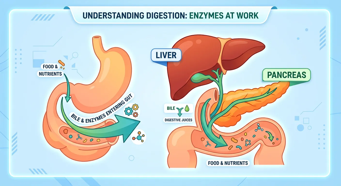
“In the small intestine, the real magic happens! The liver provides bile to break down fats, while the pancreas sends powerful enzymes to complete digestion. This is where nutrients are finally liberated from the food, ready to be absorbed into your bloodstream to power your brain and muscles.”
The small intestine is the longest part of the alimentary canal, divided into the C-shaped duodenum, a long coiled middle portion called the jejunum, and a highly coiled ileum. This is the site where the final stages of digestion occur. Chyme enters the duodenum, where it encounters bile from the liver and pancreatic juice from the pancreas via the common hepato-pancreatic duct. This duct's opening is strictly guarded by the Sphincter of Oddi. The liver, the largest gland in the body, secretes bile which is stored and concentrated in the gallbladder.
Bile contains bile pigments (bilirubin and biliverdin), bile salts, cholesterol, and phospholipids but, crucially, no enzymes. Its primary role is the emulsification of fats—breaking down large fat globules into very small micelles, which increases the surface area for lipase action. Pancreatic juice contains several inactive enzymes: trypsinogen, chymotrypsinogen, procarboxypeptidases, amylases, lipases, and nucleases. Trypsinogen is activated by an enzyme called enterokinase, secreted by the intestinal mucosa, into active trypsin. Trypsin then activates the other enzymes in the pancreatic juice.
The intestinal mucosal epithelium also contains goblet cells which secrete mucus. The secretions of the brush border cells of the mucosa along with the secretions of the goblet cells constitute the intestinal juice or 'succus entericus.' This juice contains a variety of enzymes like dipeptidases, maltase, lactase, sucrase, and nucleosidases. These enzymes act on the breakdown products of the pancreatic juice, completing the digestion of carbohydrates into monosaccharides, proteins into amino acids, and fats into fatty acids and glycerol. This chemical nexus ensures that nutrients are fully liberated and ready for absorption.
Quick Revision Points
- Bile emulsifies fats and activates lipases; it contains no enzymes.
- Pancreatic enzymes are secreted as proenzymes; Trypsinogen is activated by Enterokinase.
- Sphincter of Oddi controls the entry of bile and pancreatic juice into the duodenum.
- Succus entericus contains the 'final' enzymes for monomer production.
NEET Exam Angle
- Identify the activation cascade: Enterokinase → Trypsinogen → Trypsin → Chymotrypsinogen.
- Remember that Brunner's glands (duodenum) secrete mucus to neutralize acidic chyme.
| Enzyme | Target | Final Product |
|---|---|---|
| Trypsin | Proteins | Peptides/Amino Acids |
| Pancreatic Amylase | Polysaccharides | Disaccharides |
| Nucleases | Nucleic Acids | Nucleotides/Nucleosides |
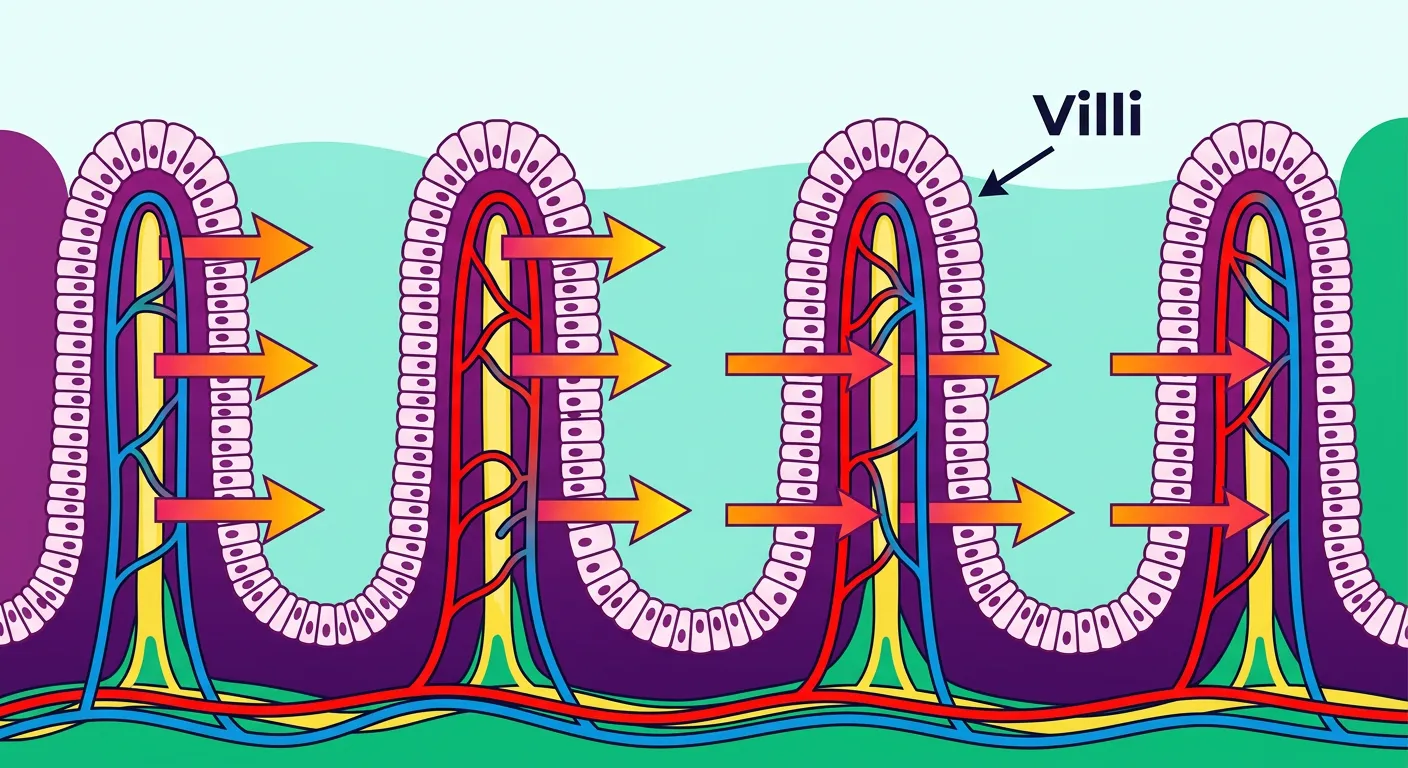
06Intestinal Villi: Structural Adaptations for Maximum Nutrient Absorption
“How does food enter the blood? Meet the villi! These tiny, finger-like projections increase the surface area of the intestine, acting like millions of microscopic vacuums soaking up glucose, amino acids, and fats. The more surface area, the faster you get the energy you need for NEET prep!”
Now that digestion is complete, the resulting simple substances are absorbed in the jejunum and ileum regions of the small intestine. The inner lining of the small intestine is folded into thousands of finger-like projections called villi. Each villus is lined by cells that possess microscopic projections called microvilli, which give a brush border appearance. This evolutionary adaptation increases the surface area for absorption exponentially—by nearly 600 times—ensuring that even a small length of intestine can process vast amounts of nutrients efficiently.
Absorption utilizes various transport mechanisms: simple diffusion, facilitated transport, and active transport. Small amounts of monosaccharides like glucose, amino acids, and some electrolytes like chloride ions are generally absorbed by simple diffusion. However, substances like fructose and some amino acids are absorbed with the help of carrier proteins, a process called facilitated transport. Nutrients like glucose and amino acids are also absorbed via active transport, which occurs against the concentration gradient and requires energy (ATP). This ensures that almost all available nutrients are reclaimed from the lumen.
Fatty acids and glycerol, being insoluble in water, cannot be absorbed directly into the blood. They are first incorporated into small droplets called micelles which move into the intestinal mucosa. There, they are re-formed into very small protein-coated fat globules called chylomicrons which are transported into the lymph vessels (lacteals) in the villi. These lymph vessels eventually release the absorbed substances into the blood stream. This distinct pathway is a classic NEET favorite, highlighting how the body manages complex nutrient transport based on chemical solubility. The efficiency of this system is the reason why we can extract sufficient energy for daily life from our meals.
Quick Revision Points
- Villi and microvilli (brush border) dramatically increase surface area for absorption.
- Active transport is used for glucose and Na+ (requires energy/ATP).
- Lacteals are specialized lymph vessels for the absorption of chylomicrons (fats).
- Micelles are small water-soluble droplets that facilitate fat entry into mucosal cells.
NEET Exam Angle
- Distinguish between the transport of glucose (active/facilitated) vs. fructose (facilitated only).
- Understand that chylomicrons are formed within the intestinal mucosal cells, not the lumen.
| Nutrient Type | Transport Path | Mechanism |
|---|---|---|
| Glucose/Amino Acids | Blood Capillaries | Active/Facilitated/Diffusion |
| Fructose | Blood Capillaries | Facilitated Diffusion |
| Fatty Acids (Chylomicrons) | Lacteals (Lymph) | Exocytosis into Lymph |
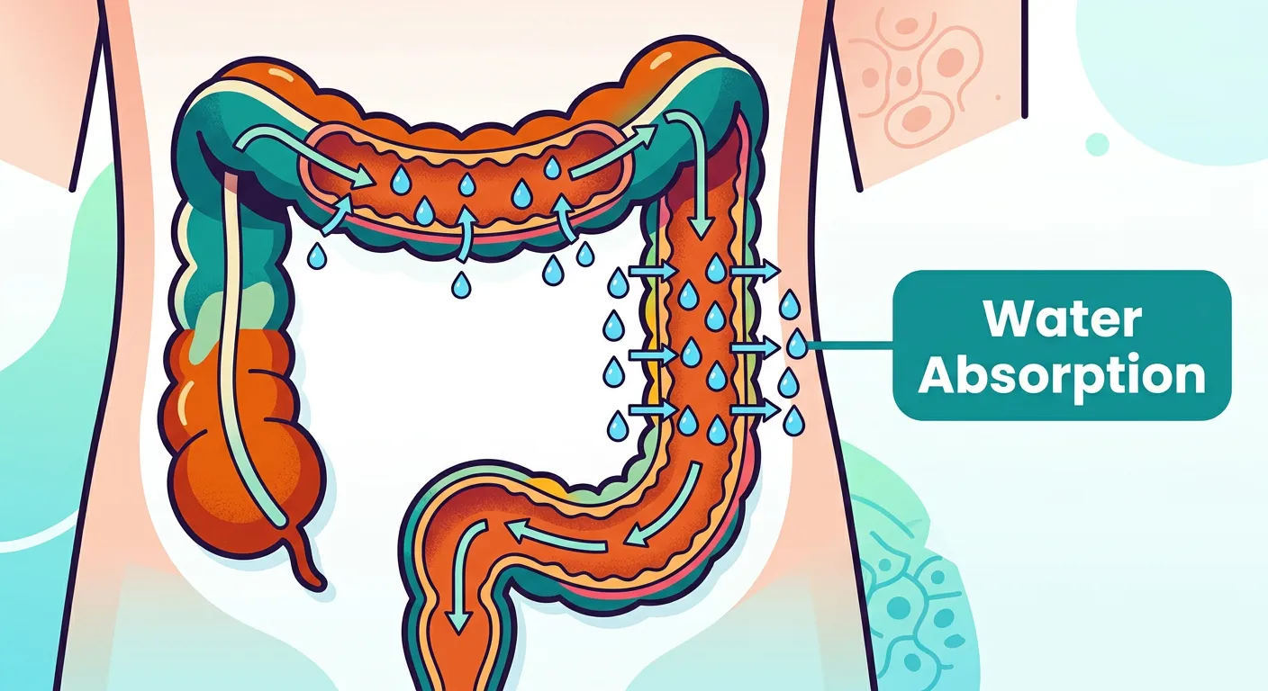
07The Large Intestine: Water Homeostasis and Waste Management
“Finally, we reach the large intestine. It doesn't digest food, but it is a master of recycling—it absorbs water and essential salts from the remaining waste. What’s left is compacted and expelled. You've just completed a full circuit of one of the body's most vital systems. Great work!”
The journey concludes in the large intestine, which is wider but shorter than the small intestine. It consists of the caecum, colon, and rectum. The caecum is a small blind sac which hosts some symbiotic micro-organisms. A narrow finger-like tubular projection, the vermiform appendix, which is a vestigial organ, arises from the caecum. The caecum opens into the colon, which is divided into four parts: an ascending, a transverse, a descending part, and a sigmoid colon. The descending part opens into the rectum which finally opens out through the anus.
No significant digestive activity occurs in the large intestine. Its primary mission is the absorption of some water, minerals, and certain drugs. It also secretes mucus which helps in adhering the waste (undigested) particles together and lubricating them for an easy passage. The undigested, unabsorbed substances called feces enter into the caecum through the ileo-caecal valve, which prevents the backflow of the fecal matter. Feces are stored in the rectum until defecation. The activities of the GI tract are under neural and hormonal control for proper coordination of different parts.
Finally, we must consider the disorders of the digestive system. Inflammation of the intestinal tract is a common ailment caused by bacterial or viral infections. Common disorders include Jaundice (liver affected, skin/eyes turn yellow due to bile pigments), Vomiting (ejection of stomach contents through the mouth), Diarrhea (increased liquidity of fecal discharge), Constipation (feces retained within the colon), and Indigestion (food not properly digested leading to a feeling of fullness). Mastery of these clinical aspects is essential for NEET, as it connects basic anatomy to real-world medical pathology, providing a complete picture of human health.
Quick Revision Points
- Large intestine regions: Caecum, Colon (4 parts), Rectum, and Anus.
- The ileo-caecal valve is a structural barrier preventing waste backflow.
- Primary function: Water and mineral absorption and waste compaction.
- Digestive disorders: Jaundice, Vomiting, Diarrhea, Constipation, Indigestion.
NEET Exam Angle
- Know the parts of the colon: Ascending, Transverse, Descending, and Sigmoid.
- Understand that the large intestine absorbs water but does not secrete digestive enzymes.
| Condition | Symptom/Cause |
|---|---|
| Jaundice | Bile pigment accumulation in tissues |
| Diarrhea | Reduced absorption of water from feces |
| Indigestion | Inadequate enzyme secretion or spicy food |
Recommended Reading
Explore related Biology topics to build deeper chapter connections for NEET.
- Morphology and Modifications · Topic 2.1
- Families · Topic 2.10
- Animal Tissues · Topic 2.11
- Frog Morphology · Topic 2.12
- Circulatory System · Topic 2.14
- Respiratory System · Topic 2.15
- Jump to Key Terms (Quick Revision)
- Review Common NEET Mistakes
- Read Topic FAQs
- Check PYQ Pattern Notes
- Practice NEET MCQs
- Solve NEET PYQs
📚 Key Terms
⚠️ Common NEET Mistakes
- 1Assuming digestion occurs in the esophagus (it is purely a transit tube).
- 2Thinking bile contains enzymes (it only emulsifies fats and activates lipases).
- 3Misidentifying the activation site of pancreatic enzymes (they are activated in the duodenum, not the pancreas).
- 4Confusing the transport of glucose (blood) and fats (lymph/lacteals).
- 5Forgetting that the dental formula for milk teeth (2102) is different from permanent teeth (2123).
- 6Assuming absorption occurs only in the small intestine (some drugs and water are absorbed in the stomach and large intestine).
📝 NEET PYQ Pattern
NEET questions frequently target the cellular sources of gastric secretions (Oxyntic vs Peptic cells), the enzyme activation sequence in the duodenum (the role of Enterokinase), and the specific mechanisms of absorption (Active vs Passive vs Facilitated). Questions on the histological layers of the GI tract and the dental formula are also recurring themes in the last decade.
❓ Frequently Asked Questions
What is the pH required for pepsin activation in the stomach?
Pepsin activation occurs at a highly acidic pH of approximately 1.8, facilitated by the secretion of Hydrochloric acid (HCl) by the parietal (oxyntic) cells.
How does salivary amylase contribute to the sweet taste of bread?
Salivary amylase breaks down complex starch molecules (polysaccharides) in bread into simpler disaccharides like maltose. These simple sugars stimulate taste receptors, creating a sweet perception.
What is the primary role of the Sphincter of Oddi in digestion?
The Sphincter of Oddi regulates the flow of bile from the common bile duct and pancreatic juices from the pancreatic duct into the duodenum, ensuring they enter only when chyme is present.
Why is the small intestine the main site for nutrient absorption compared to the stomach?
The small intestine contains specialized structures called villi and microvilli, which create an enormous surface area for absorption. Additionally, it contains the final enzymes needed to break down food into absorbable monomers.
What are lacteals, and why are they necessary for fat absorption?
Lacteals are specialized lymphatic capillaries located within the villi. They are necessary because fat molecules, repackaged as chylomicrons, are too large and water-insoluble to directly enter the narrow blood capillaries.
Does the large intestine play any role in the chemical digestion of proteins?
No, the large intestine does not secrete any digestive enzymes for protein digestion. Its primary role is restricted to water and mineral absorption, mucus secretion, and waste compaction.
Written By
NEET Content Strategist & Biology Expert
Sangita Kumari is a NEET educator and content strategist with over 6 years of experience teaching Biology, Chemistry, and Physics to Class 11 and 12 aspirants. She helps bridge the gap between traditional NCERT preparation and modern AI-powered learning. Her content is trusted by thousands of NEET aspirants across India.