🎬 Video Lesson Available
Watch the full 7-slide video lesson for Circulatory System with AI teacher narration and visual explanations.
01The Human Circulatory System: The Body's Ultimate Courier Network
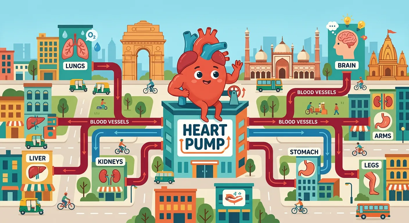
“Welcome, NEET warriors! Think of your circulatory system as the ultimate courier service of your body. Just like a busy delivery network, it ensures oxygen and nutrients reach every single cell, while clearing out the trash. Today, we decode the heart, the engine behind it all!”
In the complex ecosystem of the human body, the circulatory system acts as the lifeline that connects every cell, tissue, and organ. Think of it as a sophisticated, high-speed logistics network. While our respiratory system brings in oxygen and our digestive system absorbs nutrients, these resources are useless unless they reach their final destination: the cells. This is where the circulatory system steps in. It functions as a closed-loop transportation hub, ensuring that oxygen-rich blood is delivered precisely where it's needed while simultaneously acting as a waste management service. By hauling away metabolic byproducts like carbon dioxide and urea, the system prevents toxic buildup, maintaining the delicate balance of homeostasis.
The centerpiece of this entire operation is the heart, a muscular pump that never takes a break. It generates the hydrostatic pressure required to propel blood through miles of vascular tubing. For a NEET aspirant, it is crucial to view this system not just as a set of pipes, but as a dynamic regulatory mechanism. Efficient circulation is the reason endothermic organisms like humans can maintain a constant body temperature and support high metabolic activities. Without this rapid delivery and pickup service, our cells would essentially suffocate in their own waste within minutes. Understanding the circulatory system is the foundation for mastering animal physiology, as it interacts directly with the excretory, respiratory, and endocrine systems.
| Component | Primary Role in Transport | Substance Handled |
|---|---|---|
| Arterial Blood | Distribution | Oxygen, Glucose, Hormones |
| Venous Blood | Collection | Carbon Dioxide, Metabolic Waste |
| Plasma | Solvent Medium | Water, Ions, Proteins, Urea |
Quick Revision Points
- The human circulatory system is a 'closed' type, meaning blood remains within vessels.
- It serves as the primary medium for transporting gases (O2/CO2) and nutrients.
- Homeostasis is heavily dependent on the regulation of blood flow and pressure.
- The heart provides the mechanical energy needed to overcome vascular resistance.
- Waste management (removing CO2) is just as critical as nutrient delivery.
NEET Exam Angle
- Focus on the distinction between 'Open' and 'Closed' circulatory systems (e.g., Arthropods vs. Chordates).
- Note that in humans, the system is strictly closed, which allows for higher blood pressure and more efficient delivery.
- Questions often link circulation to the Excretory system regarding the transport of nitrogenous wastes to the kidneys.
02Anatomy of the Heart: Understanding the Four-Chambered Masterpiece
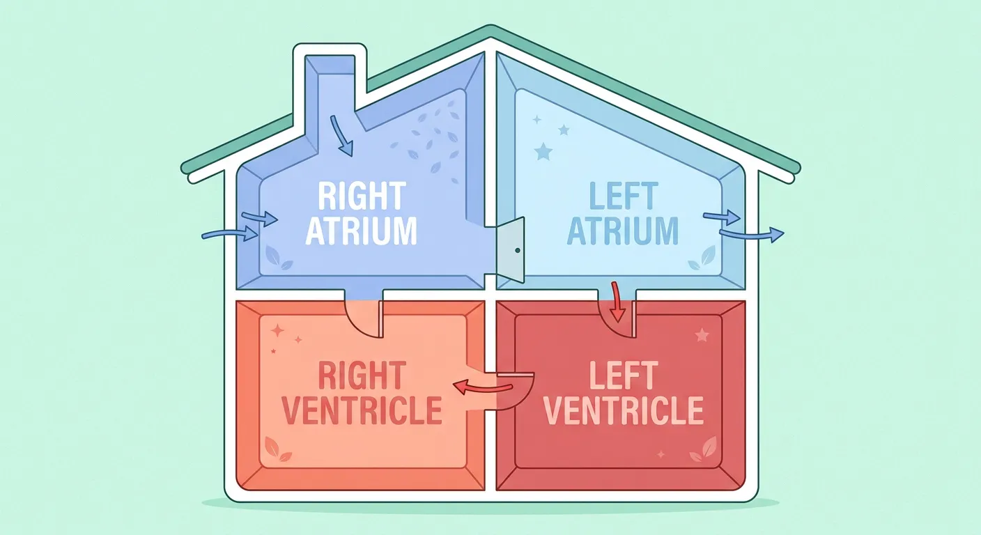
“Meet the four-chambered heart! Think of it as a duplex house: two upper rooms called Atria, and two sturdy lower rooms called Ventricles. The right side handles deoxygenated blood, while the left side pumps fresh, oxygen-rich blood. It is a masterpiece of biological engineering!”
The human heart is a marvel of evolutionary engineering, specifically designed to prevent the mixing of oxygenated and deoxygenated blood. Located in the thoracic cavity within the mediastinum, it is roughly the size of a clenched fist. The heart is divided into four distinct chambers: two superior, thin-walled Atria and two inferior, thick-walled Ventricles. The Atria act as receiving chambers, collecting blood returning from the body (Right Atrium) and the lungs (Left Atrium). Their walls are relatively thin because they only need to push blood into the ventricles directly below them, a task assisted by gravity and a small pressure gradient.
In contrast, the Ventricles are the 'powerhouses' of the heart. The Right Ventricle pumps deoxygenated blood to the lungs via the pulmonary artery—a relatively short distance. However, the Left Ventricle is the most muscular chamber of all. Its myocardium is significantly thicker because it must generate enough force to eject blood into the aorta and throughout the entire systemic circuit, reaching everything from the scalp to the toes. This structural difference is a classic example of 'form following function.' The four-chambered design is a significant evolutionary leap from the two-chambered hearts of fish or three-chambered hearts of reptiles, providing the high-pressure oxygen delivery required for the high metabolic demands of mammals.
| Chamber | Wall Thickness | Blood Type | Destination |
|---|---|---|---|
| Right Atrium | Thin | Deoxygenated | Right Ventricle |
| Left Atrium | Thin | Oxygenated | Left Ventricle |
| Right Ventricle | Medium | Deoxygenated | Lungs (Pulmonary) |
| Left Ventricle | Thickest | Oxygenated | Whole Body (Systemic) |
Quick Revision Points
- The heart is protected by a double-layered membrane called the pericardium.
- The Interatrial Septum separates the two atria, while the Interventricular Septum separates the ventricles.
- Left ventricular walls are roughly 3 times thicker than right ventricular walls.
- Oxygenated blood is strictly confined to the left side of the heart in healthy individuals.
- Deoxygenated blood is strictly confined to the right side of the heart.
NEET Exam Angle
- Remember the sequence: Right Atrium -> Right Ventricle -> Lungs -> Left Atrium -> Left Ventricle.
- NEET often asks about the 'thickest wall'—it is always the Left Ventricle.
- Understand the evolutionary advantage: Four chambers allow for complete separation of blood, preventing 'mixing' which increases efficiency.
03Hematology Basics: Red Blood Cells as Essential Oxygen Couriers
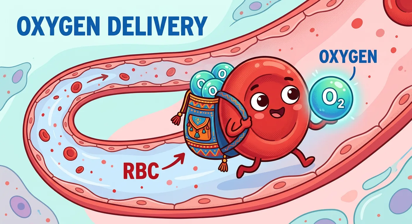
“Blood is not just red liquid; it is a fleet of delivery vehicles! Red blood cells are your hardworking couriers, carrying oxygen to every corner of your body. They are essential for life, acting just like a rapid delivery app ensuring your cells never run out of fuel.”
Blood is often called the 'fluid of life,' and its most iconic component is the Erythrocyte, or Red Blood Cell (RBC). These cells are specialized for one primary mission: gas transport. In mature mammals, RBCs undergo a unique transformation where they lose their nucleus, mitochondria, and other organelles. This process, known as enucleation, creates more internal space for Hemoglobin, the iron-containing pigment that binds to oxygen. The biconcave shape of an RBC isn't just for show; it maximizes the surface-area-to-volume ratio, facilitating rapid diffusion of oxygen into and out of the cell. Furthermore, the lack of mitochondria ensures that the RBC doesn't consume the oxygen it is meant to deliver, relying instead on anaerobic metabolism.
The lifespan of an RBC is approximately 120 days, after which they are filtered out and destroyed in the spleen, often referred to as the 'graveyard of RBCs.' A healthy adult has roughly 5 to 5.5 million RBCs per cubic millimeter of blood. This density is not static; it can adapt to environmental changes. For instance, people living at high altitudes produce more RBCs to compensate for lower oxygen levels in the air, a process regulated by the hormone erythropoietin from the kidneys. For NEET, understanding the morphology of RBCs is vital, especially how their flexibility allows them to squeeze through capillaries that are narrower than the cells themselves.
Quick Revision Points
- Erythrocytes are biconcave and disc-shaped to increase surface area.
- Mature mammalian RBCs lack a nucleus and most organelles (for more Hemoglobin space).
- Hemoglobin consists of four globin chains, each with a central 'heme' group containing iron.
- RBCs are produced in the red bone marrow through a process called erythropoiesis.
- The spleen is the site where aged or damaged RBCs are recycled.
NEET Exam Angle
- Frequent question: Why do RBCs lack mitochondria? Answer: To prevent the consumption of oxygen during transport.
- Note the role of iron: Iron deficiency leads to anemia, reducing the blood's oxygen-carrying capacity.
- High-altitude adaptation (Polycythemia) is a common topic in 'Respiration' and 'Circulation' crossover questions.
- Remember: RBCs are the most numerous of all blood cells.
04Vascular Highways: Arteries, Veins, and the Capillary Exchange
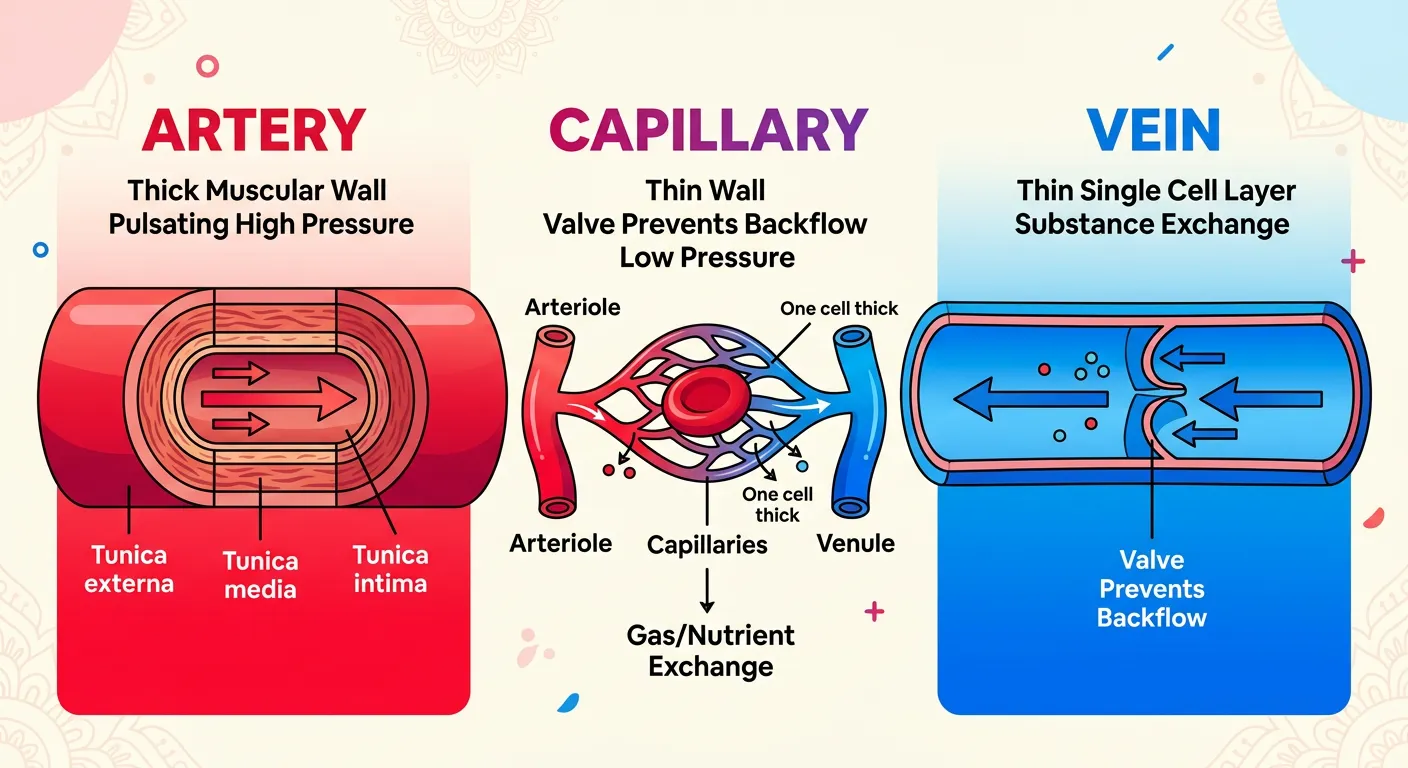
“Your blood vessels are the highway system. Arteries are like high-speed expressways taking blood away from the heart, while Veins are the return lanes bringing it back. Capillaries? They are the narrow service lanes where the actual exchange of gases and nutrients happens with your cells.”
To move blood throughout the body, the heart relies on a diverse network of vessels. Arteries are the high-pressure 'expressways' that carry blood away from the heart. To withstand the surge of blood during ventricular contraction, arteries have thick, elastic walls. Conversely, Veins are the 'return lanes' that bring blood back to the heart. Because blood pressure in the veins is significantly lower, their walls are thinner, and they possess one-way valves. These valves are critical because they prevent the backflow of blood, especially in the limbs where blood must travel against gravity to return to the heart.
Between the massive arteries and veins lies the most critical part of the network: the Capillaries. These are microscopic vessels with walls only one cell layer thick (tunica intima). This extreme thinness allows for the actual 'exchange' to happen. Oxygen and nutrients diffuse out into the tissues, while carbon dioxide and wastes diffuse into the blood. Structurally, all large vessels (except capillaries) consist of three layers: the Tunica Intima (inner endothelium), the Tunica Media (middle layer of smooth muscle and elastic fibers), and the Tunica Adventitia/Externa (outer layer of connective tissue). The Tunica Media is notably thicker in arteries to accommodate the pulsating pressure from the heart.
| Vessel Type | Wall Structure | Pressure | Presence of Valves |
|---|---|---|---|
| Artery | Thick Tunica Media | High | Absent (except at base of aorta/pulmonary artery) |
| Vein | Thin Tunica Media | Low | Present (prevent backflow) |
| Capillary | Single cell layer (Intima) | Very Low | Absent |
Quick Revision Points
- Arteries generally carry oxygenated blood (except the Pulmonary Artery).
- Veins generally carry deoxygenated blood (except the Pulmonary Vein).
- Capillaries have the slowest blood flow to allow sufficient time for exchange.
- Tunica Media is made of smooth muscle and is under the control of the autonomic nervous system.
- Veins have a larger lumen (internal space) compared to arteries.
NEET Exam Angle
- Comparison of Tunica Media is a high-yield point—it's always thicker in arteries.
- Understand the role of valves in veins: Skeletal muscle contraction 'milks' blood toward the heart (the skeletal muscle pump).
- Be careful: Not all arteries carry oxygenated blood; 'Artery' is defined by direction (away from heart), not oxygen status.
05The Mechanism of Double Circulation: Pulmonary and Systemic Circuits
“Here is the NEET magic: Double Circulation! Blood passes through your heart twice in one complete round. First, it goes to the lungs for a fresh oxygen refill, then it journeys to the rest of the body. It is efficient, organized, and absolutely essential for us humans.”
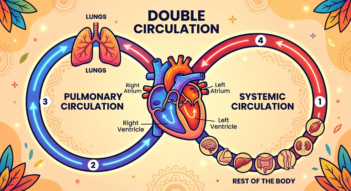
Humans utilize a system known as 'Double Circulation,' which means blood passes through the heart twice during one complete cycle through the body. This is divided into two distinct circuits: Pulmonary and Systemic. Pulmonary circulation is the shorter loop. It begins in the Right Ventricle, which pumps deoxygenated blood through the Pulmonary Artery to the lungs. Here, CO2 is exchanged for O2. The now oxygenated blood returns to the Left Atrium via the Pulmonary Veins. This circuit's primary purpose is strictly gas exchange—recharging the blood with fresh oxygen.
Systemic circulation is the more extensive loop. It starts in the Left Ventricle, which pumps the oxygen-rich blood through the Aorta to the rest of the body. After delivering oxygen and picking up waste at the capillary level, the now deoxygenated blood returns to the Right Atrium through the Vena Cava (Superior and Inferior). This dual-circuit system is incredibly efficient because it ensures that tissues always receive the most oxygenated blood possible. It prevents the dilution of oxygenated blood with deoxygenated blood, a flaw seen in animals with three-chambered hearts. This separation is what allows humans to maintain high metabolic rates and constant internal temperatures, regardless of the environment.
| Circuit | Originating Chamber | Destination | Returning Chamber |
|---|---|---|---|
| Pulmonary | Right Ventricle | Lungs | Left Atrium |
| Systemic | Left Ventricle | Tissues/Body | Right Atrium |
Quick Revision Points
- Double circulation involves two separate pathways: Pulmonary and Systemic.
- The Pulmonary Artery is the only artery carrying deoxygenated blood.
- The Pulmonary Vein is the only vein carrying oxygenated blood.
- Portal systems (like the Hepatic Portal System) are specialized additions to systemic circulation.
- This system is essential for endothermy (warm-bloodedness).
NEET Exam Angle
- Flowcharts of double circulation are extremely common in exams. Practice tracing a molecule of CO2 from the toe to the lung.
- Understand the Hepatic Portal System: Blood from the digestive tract goes to the liver before returning to the heart.
- Why double circulation? To provide a high-pressure system for systemic delivery while maintaining a lower pressure for delicate lung tissues.
06The Cardiac Cycle: Valve Mechanics and the 'Lub-Dub' Heart Sounds

“Ever wondered about that 'Lub-Dub' sound? It is the music of your heart valves closing! The 'Lub' happens when the AV valves shut, and the 'Dub' follows when the semilunar valves snap closed. It is the rhythmic heartbeat that keeps you alive and thriving every second.”
The rhythm of the heart, known as the cardiac cycle, is a sequence of events that occurs during one heartbeat. To ensure blood flows in only one direction, the heart uses a set of four valves. These valves don't open or close on their own; they respond to pressure changes within the chambers. The 'Lub-Dub' sound you hear through a stethoscope is the sound of these valves slamming shut. The first sound, 'Lub' (S1), occurs when the Atrioventricular (AV) valves—the Tricuspid and Bicuspid (Mitral) valves—close. This happens at the beginning of ventricular systole (contraction) as the pressure in the ventricles rises sharply, forcing the valves shut to prevent blood from flowing back into the atria.
The second sound, 'Dub' (S2), occurs when the Semilunar valves (at the base of the Aorta and Pulmonary Artery) close. This happens at the beginning of ventricular diastole (relaxation) as the ventricles begin to refill and the pressure within them drops. The back-pressure from the arteries snaps the semilunar valves shut to prevent blood from leaking back into the ventricles. Any abnormality in these sounds, such as a 'whooshing' or 'murmur,' often indicates a valvular defect where a valve is either too narrow (stenosis) or leaking (regurgitation). For NEET, it is crucial to link these sounds to specific phases of the cardiac cycle.
| Sound | Valve Involved | Phase of Cardiac Cycle | Significance |
|---|---|---|---|
| 'Lub' (S1) | Tricuspid & Bicuspid | Start of Ventricular Systole | Closure of AV valves |
| 'Dub' (S2) | Semilunar Valves | Start of Ventricular Diastole | Closure of Semilunar valves |
Quick Revision Points
- Valves ensure unidirectional (one-way) blood flow.
- Tricuspid valve: Between Right Atrium and Right Ventricle.
- Bicuspid (Mitral) valve: Between Left Atrium and Left Ventricle.
- The cardiac cycle lasts approximately 0.8 seconds in a resting human.
- Heart sounds are heard using a stethoscope; 'Lub' is louder and longer than 'Dub'.
NEET Exam Angle
- Match the valve closure to the sound: AV valves = Lub; Semilunar = Dub.
- Be clear on the timing: Valves close when pressure in the following chamber/vessel becomes higher than the originating chamber.
- Questions may ask about the state of the atria during the 'Lub' sound (they are in diastole).
07NEET Strategy: Mastering the Rhythm of Life and Exam High-Yields
“You have mastered the basics! From the heart's four chambers to the pulse in your veins, you now understand the rhythm of life. Keep this flow chart in mind, stay curious, and keep smashing those NEET goals. Your dream college is just one concept away!”
As we wrap up the Circulatory System, your goal as a NEET aspirant is to integrate these concepts into a functional mental map. Don't just memorize the names; understand the 'Why.' Why is the left ventricle thicker? To overcome systemic resistance. Why do veins have valves? To prevent backflow under low pressure. A common confusion point to watch out for is the oxygenation status of the pulmonary vessels. Always remember: Pulmonary Arteries take deoxygenated blood to the lungs, and Pulmonary Veins bring oxygenated blood back. This is the reverse of the general rule for systemic vessels.
Consistency in revision is your best friend. Draw the double circulation flowchart until you can do it from memory. Pay close attention to the specific timings of the cardiac cycle (Atrial systole: 0.1s, Ventricular systole: 0.3s, Joint diastole: 0.4s). Use mnemonics for the valves—'Tricuspid is on the Right' (both have an 'R'). By mastering these high-yield topics, you aren't just preparing for an exam; you're learning the fundamental mechanics of human life. Keep your focus sharp, stay curious, and remember that every concept you master brings you one step closer to your medical career.
Quick Revision Flowchart
- Right Atrium receives CO2-rich blood from Vena Cava.
- Through Tricuspid Valve to Right Ventricle.
- Through Pulmonary Valve to Pulmonary Artery to Lungs.
- O2-rich blood returns via Pulmonary Veins to Left Atrium.
- Through Bicuspid Valve to Left Ventricle.
- Through Aortic Valve to Aorta to Body Tissues.
NEET Exam Angle
- The Cardiac Output formula (Stroke Volume x Heart Rate) is frequently tested. Normal is approx. 5 Liters/min.
- Focus on the nodal tissue (SA Node, AV Node, Bundle of His, Purkinje fibers) as the electrical 'spark' of the cycle.
- Be prepared for 'Assertion-Reason' type questions regarding the structural differences between arteries and veins.
- Keep an eye on clinical disorders like Hypertension and CAD (Coronary Artery Disease) which often appear in the same chapter.
Recommended Reading
Explore related Biology topics to build deeper chapter connections for NEET.
- Morphology and Modifications · Topic 2.1
- Families · Topic 2.10
- Animal Tissues · Topic 2.11
- Frog Morphology · Topic 2.12
- Digestive System · Topic 2.13
- Respiratory System · Topic 2.15
- Jump to Key Terms (Quick Revision)
- Review Common NEET Mistakes
- Read Topic FAQs
- Check PYQ Pattern Notes
- Practice NEET MCQs
- Solve NEET PYQs
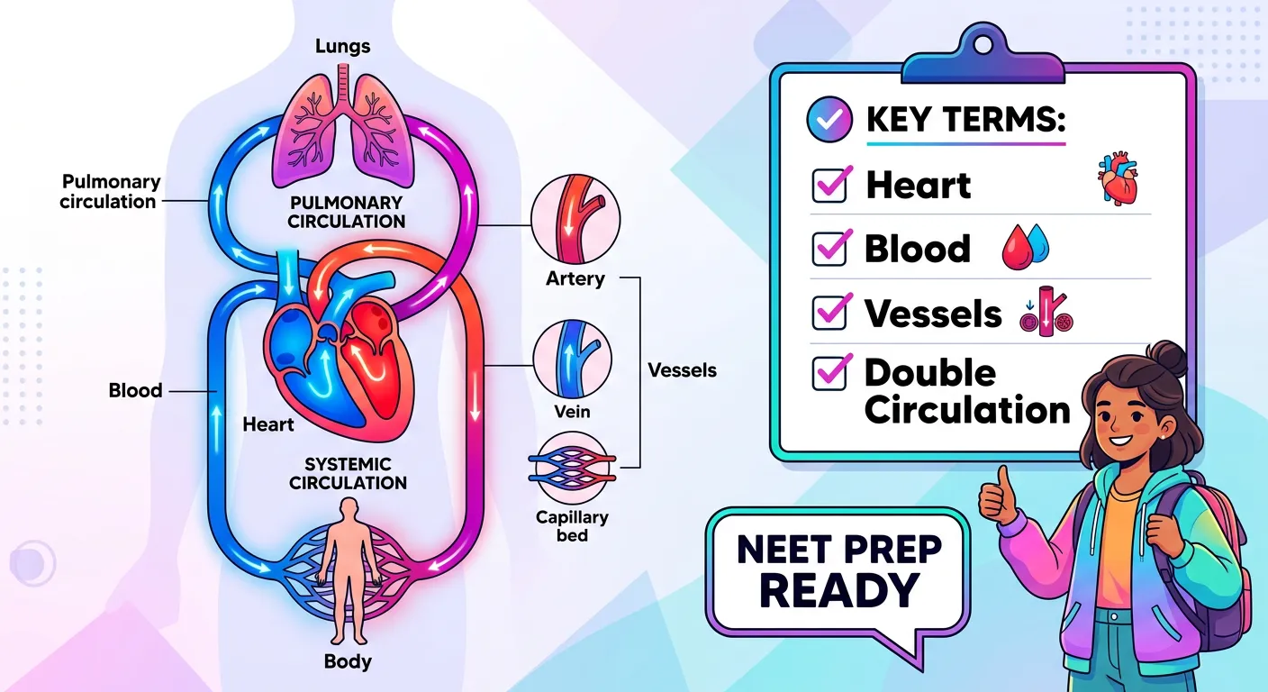
📚 Key Terms
⚠️ Common NEET Mistakes
- 1Thinking that all arteries carry oxygenated blood; remember the Pulmonary Artery is the exception.
- 2Confusing the location of the Bicuspid (Left) and Tricuspid (Right) valves.
- 3Assuming the heart sound is made by the heart muscle contracting, rather than the valves closing.
- 4Believing that mature mammalian RBCs have a nucleus; they lose it to make room for hemoglobin.
- 5Incorrectly identifying the Pulmonary Vein as a carrier of deoxygenated blood because it is a 'vein'.
📝 NEET PYQ Pattern
Questions on the human circulatory system in NEET (2018–2024) frequently focus on the sequence of blood flow through the double circulation path and the specific valve actions during the cardiac cycle. Identifying the correct oxygenation status of pulmonary vessels remains a recurring high-yield area.
❓ Frequently Asked Questions
Why is the human circulatory system called 'Double Circulation'?
It is called double circulation because blood passes through the heart twice for every one complete circuit of the body. This involves two distinct loops: the Pulmonary circuit (heart to lungs and back) and the Systemic circuit (heart to rest of the body and back).
What is the specific cause of the 'Lub' and 'Dub' heart sounds?
The 'Lub' sound (S1) is caused by the closure of the Atrioventricular valves (Tricuspid and Bicuspid) at the start of ventricular systole. The 'Dub' sound (S2) is caused by the closure of the Semilunar valves at the start of ventricular diastole.
How do arteries differ structurally from veins to handle higher blood pressure?
Arteries have a significantly thicker and more elastic Tunica Media (middle layer) compared to veins. This muscular wall allows them to withstand and regulate the high pressure of blood being ejected from the ventricles.
Which heart chamber is responsible for pumping oxygenated blood to the whole body?
The Left Ventricle is responsible for pumping oxygenated blood into the aorta and throughout the systemic circulation. This is why it has the thickest muscular walls of all four heart chambers.
What is the role of valves in the veins and the heart?
Valves act as one-way gates that ensure blood flows in only one direction and prevent the backflow of blood. In the heart, they prevent blood from flowing back into previous chambers; in veins, they prevent blood from pooling due to gravity.
Why are capillaries the only vessels where gas exchange occurs?
Capillaries are the only vessels thin enough for exchange because their walls consist of only a single layer of squamous epithelial cells (endothelium). This minimizes the distance gases and nutrients must diffuse between the blood and the tissues.
Written By
NEET Content Strategist & Biology Expert
Sangita Kumari is a NEET educator and content strategist with over 6 years of experience teaching Biology, Chemistry, and Physics to Class 11 and 12 aspirants. She helps bridge the gap between traditional NCERT preparation and modern AI-powered learning. Her content is trusted by thousands of NEET aspirants across India.