🎬 Video Lesson Available
Watch the full 7-slide video lesson for Nervous System with AI teacher narration and visual explanations.
01Architecture of the Human Neural System: CNS and PNS Explained
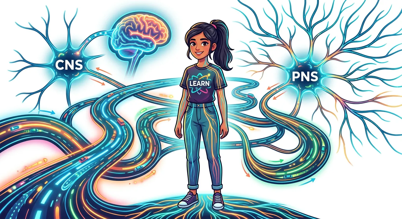
“Welcome! Think of your nervous system as the ultimate superhighway of your body. Just like the Delhi Metro connects every corner of the city, your nervous system coordinates everything you do. It is split into the Central Nervous System, your command center, and the Peripheral Nervous System, the messengers.”
The human nervous system is an engineering marvel, designed to integrate, process, and respond to both internal and external stimuli with incredible precision. To understand how we perceive the world, we must first look at the body’s organizational hierarchy-and-systematics). The neural system is primarily divided into two main components: the Central Nervous System (CNS) and the Peripheral Nervous System (PNS). The CNS serves as the primary command center where all information is evaluated and decisions are made. In contrast, the PNS acts as the expansive network of cables connecting this central hub to the rest of the body’s periphery—your skin, muscles, and organs.
Think of the CNS as the 'CPU' of your body. Comprising the brain and the spinal cord, it acts as the primary site for information processing and control. Every conscious decision, motor command, and memory storage happens here. The PNS, meanwhile, is composed of all the nerves of the body associated with the CNS. These nerves are categorized into two types of fibers: afferent (sensory) fibers, which carry impulses from tissues to the CNS, and efferent (motor) fibers, which transmit regulatory impulses from the CNS to the target tissues or organs.
Within the PNS, we further classify the system into somatic and autonomic branches. The somatic nervous system handles voluntary movements—like deciding to lift your arm—by relaying impulses from the CNS to skeletal muscles. The autonomic nervous system runs the background processes, such as digestion and heart rate regulation, by transmitting impulses to involuntary organs and smooth muscles. The autonomic system is further subdivided into the sympathetic and parasympathetic nervous systems, which often act antagonistically to maintain internal balance. Mastering this structural hierarchy is the foundational step for any serious NEET aspirant, as it sets the stage for all subsequent physiological chapters including locomotion and chemical coordination.
Quick Revision Points
- CNS includes the brain and spinal cord, serving as the command center.
- PNS consists of all nerves outside the CNS, facilitating communication via afferent and efferent fibers.
- Somatic neural system transmits impulses from the CNS to skeletal muscles (voluntary control).
- Autonomic neural system coordinates involuntary activities like smooth muscle contraction and cardiac rhythm.
- Visceral nervous system is a part of the PNS comprising the whole complex of nerves, fibers, and ganglia by which impulses travel from the CNS to the viscera.
NEET Exam Angle
- Focus on the distinction between the CNS and PNS as the organizational basis for all physiology questions.
- Identify the difference between afferent (sensory) and efferent (motor) nerve fibers clearly, as these are frequently tested in MCQ formats.
- Be prepared to differentiate between somatic (voluntary) and autonomic (involuntary) pathways in clinical test scenarios.
| Feature | CNS | PNS |
|---|---|---|
| Main Components | Brain & Spinal Cord | Cranial & Spinal Nerves |
| Primary Function | Processing & Integration | Transmission/Communication |
| Control | Voluntary/Involuntary Central Hub | Links to limbs/organs/viscera |
| Nerve Fiber Types | Not applicable (Interneurons) | Afferent and Efferent |
02The Neuron: Anatomy of the Functional Unit of the Nervous System
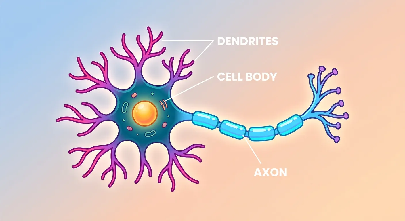
“Meet the Neuron, the superstar cell! Think of it like a text message sender. Dendrites are the antennas receiving information, the Cell Body is the processor, and the Axon is the long wire that transmits the message at lightning speed to the next cell.”
If the nervous system is a computer network, the neuron is its basic transistor. Neurons are specialized cells that transmit information via electrical impulses and chemical signals. Unlike most cells, neurons are excitable and have a unique structure adapted for rapid communication. The structure of a typical neuron features a cell body (cyton) containing a nucleus and cytoplasm packed with granular bodies known as Nissl’s granules. These granules are rich in rough endoplasmic reticulum and are essential sites for protein synthesis, ensuring the cell remains functional despite its long projections.
Branching out from the cell body are short, tree-like extensions called dendrites. These processes contain Nissl's granules and are designed to receive incoming signals and transmit them toward the cell body. In contrast, the long, slender projection known as the axon transmits impulses away from the cell body toward other neurons or effector cells. The axon ends in a branch-like structure where each branch terminates as a bulb-like 'synaptic knob,' which contains synaptic vesicles filled with neurotransmitters.
Many axons in the PNS are wrapped in a fatty myelin sheath, produced by Schwann cells. This sheath acts as an insulator, drastically increasing the speed of nerve impulse propagation. The gaps between adjacent myelin sheaths are called the Nodes of Ranvier, which are crucial for 'saltatory conduction,' where the signal essentially 'jumps' between nodes. In the CNS, oligodendrocytes perform the myelination. Structurally, neurons are classified based on the number of axons and dendrites: unipolar (found in embryonic stages), bipolar (one dendrite and one axon), and multipolar (one axon and multiple dendrites). For your NEET exams, memorizing these shapes and their specific locations—like bipolar neurons in the retina—is a classic high-yield task.
Quick Revision Points
- Cyton (cell body) contains Nissl’s granules for vital protein synthesis.
- Dendrites act as receptors, bringing impulses to the cell body.
- Axon serves as the transmitter, carrying impulses away from the cell body to the synapse.
- Myelin sheath, provided by Schwann cells or oligodendrocytes, increases transmission speed.
- Nodes of Ranvier facilitate saltatory conduction, making impulse travel significantly faster.
NEET Exam Angle
- Always recall where bipolar neurons are found: retina of the eye (highly frequently asked).
- Understand that Nissl's granules are present in the dendrites and cyton but are absent in the axon.
- Schwann cells form the myelin sheath in the PNS, while oligodendrocytes do so in the CNS.
| Neuron Type | Structural Characteristic | Common Location |
|---|---|---|
| Unipolar | One process (axon) only | Embryonic stage |
| Bipolar | One axon, one dendrite | Retina of eye, olfactory epithelium |
| Multipolar | One axon, many dendrites | Cerebral cortex (most common) |
03Synaptic Transmission: Communication via Chemical Messengers
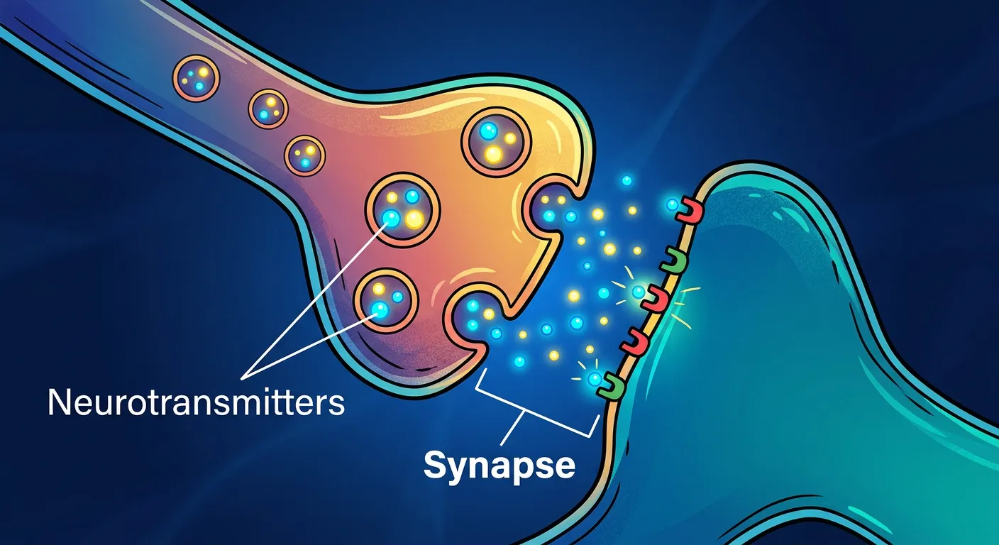
“But wait, neurons don't touch! There is a tiny gap called the Synapse. How do they communicate? They release chemical messengers called Neurotransmitters. Imagine throwing a ball across a narrow street; that chemical jump is how your brain tells your finger to move or your heart to beat.”
Communication between neurons occurs at a junction called a synapse. Since the membranes of two neurons do not physically touch, the gap—the synaptic cleft—must be bridged to ensure signal continuity. A synapse is formed by the membranes of a pre-synaptic neuron and a post-synaptic neuron. There are two main types: electrical and chemical. Electrical synapses are joined by gap junctions, allowing ions to flow directly between cells. This provides extremely fast transmission, but these are rare in the human system compared to chemical synapses.
In a chemical synapse, when an action potential (nerve impulse) reaches the axon terminal, it triggers the opening of voltage-gated calcium channels. The influx of calcium ions causes synaptic vesicles to move toward the membrane, fuse with it, and release neurotransmitters into the synaptic cleft via exocytosis. These chemicals, such as Acetylcholine or GABA, diffuse across the gap and bind to specific receptor sites on the post-synaptic membrane. This binding opens ion channels, allowing the entry of ions that can generate a new potential (either excitatory or inhibitory) in the receiving neuron.
As a student, you must understand the 'All-or-None' principle, which dictates that a neuron either fires a full-strength impulse or does not fire at all once a threshold is reached. Furthermore, the refractory period ensures that the impulse travels only in one direction. This binary, highly reliable communication ensures your brain maintains control over every delicate movement and thought process. Chemical synapses, though slower than electrical ones, allow for the massive integration of signals required for complex human behaviors, making them the dominant form of communication in our nervous system.
Quick Revision Points
- Synapse: Junction consisting of a pre-synaptic membrane, synaptic cleft, and post-synaptic membrane.
- Neurotransmitters: Chemicals like Acetylcholine that bridge the gap between neurons.
- Electrical synapse: Faster, rare, allows bidirectional flow through gap junctions.
- Chemical synapse: Slower, common, unidirectional flow via neurotransmitter release.
- Action Potential: The electrical 'spike' that travels along the axon to trigger the synapse.
NEET Exam Angle
- Be ready to describe the sequence: Axon terminal depolarization -> Calcium influx -> Vesicle fusion -> Neurotransmitter release -> Receptor binding.
- Distinguish clearly between excitatory (EPSP) and inhibitory (IPSP) post-synaptic potentials.
- Focus on the fact that transmission across a chemical synapse is always unidirectional.
| Property | Electrical Synapse | Chemical Synapse |
|---|---|---|
| Speed | Extremely fast (near instant) | Slower (synaptic delay) |
| Direct Ion Flow | Yes (via gap junctions) | No (requires chemical diffusion) |
| Ubiquity | Rare in humans | Abundant and complex |
| Direction | Can be bidirectional | Strictly Unidirectional |
04The Human Brain: Forebrain, Midbrain, and Hindbrain Specializations
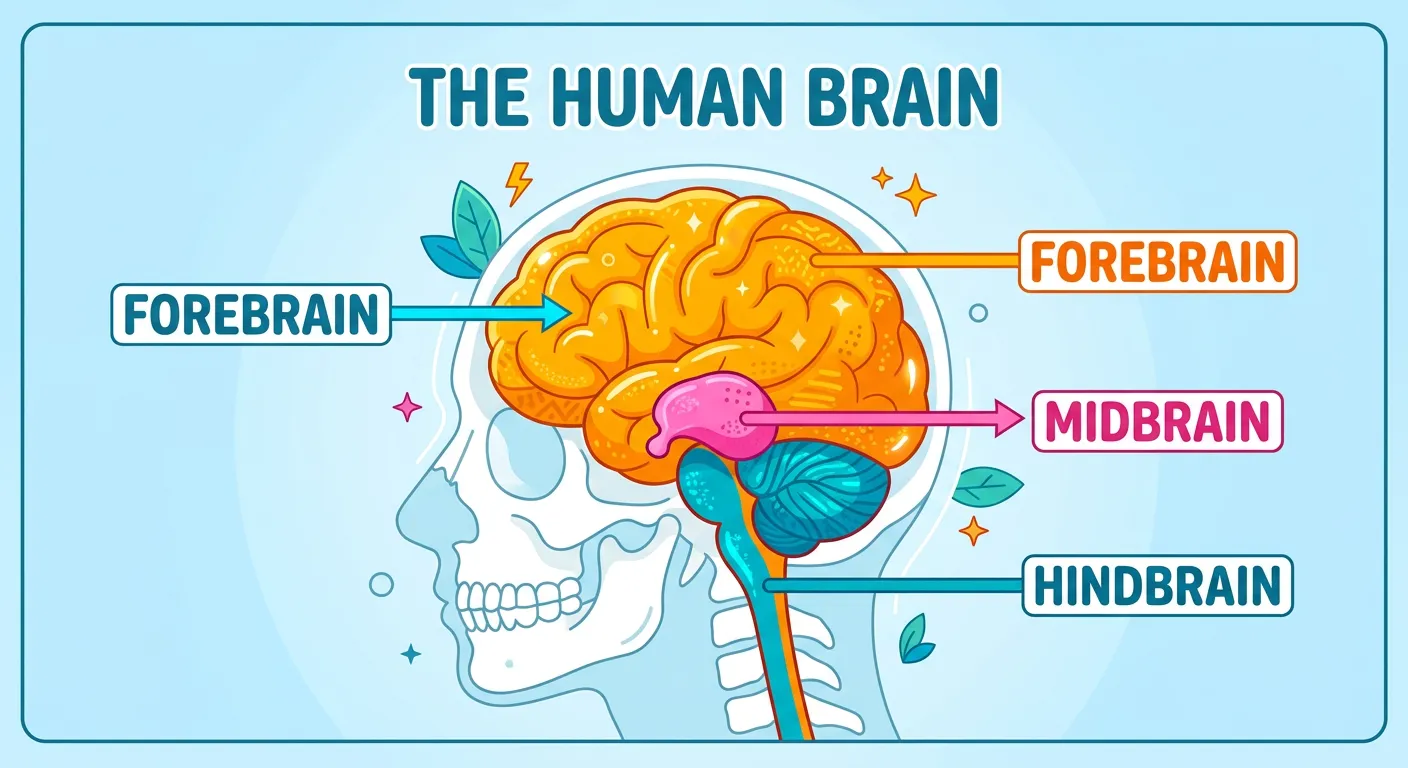
“Your brain, the Chief Minister of your body, is divided into three: the Forebrain, the smartest part for memory and logic; the Midbrain for visual and auditory reflexes; and the Hindbrain, the guardian that keeps you breathing even while you are fast asleep.”
The brain is the central information processing organ of our body and acts as the 'command and control system.' It is protected by the skull and three meningeal layers. We divide the brain into three primary parts: the forebrain, midbrain, and hindbrain. The forebrain consists of the cerebrum, thalamus, and hypothalamus. The cerebrum is the largest part, divided into two hemispheres by the corpus callosum. It contains the cerebral cortex, often called 'grey matter' due to the high concentration of neuron cell bodies. This area is responsible for high-level functions like intelligence, memory, and voluntary activity.
Deep within the forebrain, the thalamus acts as a major relay station for sensory and motor signaling. Just below it lies the hypothalamus, a vital master controller of homeostasis. It regulates body temperature, the urge for eating and drinking, and contains several groups of neurosecretory cells that secrete hormones. The forebrain also includes the limbic system (amygdala and hippocampus), which, along with the hypothalamus, regulates sexual behavior and emotional expressions like excitement, pleasure, and fear.
The midbrain is located between the thalamus of the forebrain and the pons of the hindbrain. A canal called the cerebral aqueduct passes through the midbrain. The dorsal portion of the midbrain consists of four round swellings called corpora quadrigemina, which manage visual and auditory reflexes. Finally, the hindbrain comprises the pons, cerebellum, and medulla. The cerebellum has a very convoluted surface to provide additional space for many more neurons, ensuring smooth motor coordination and balance. The medulla oblongata is connected to the spinal cord and contains centers which control respiration, cardiovascular reflexes, and gastric secretions. Damage to the medulla is often fatal because it governs these vital life processes.
Quick Revision Points
- Forebrain: Cerebrum (thought), Thalamus (relay), Hypothalamus (homeostasis/limbic system).
- Midbrain: Corpora quadrigemina (visual/auditory reflexes) and cerebral aqueduct.
- Hindbrain: Cerebellum (precision/balance), Pons (fiber tracts), Medulla (vital involuntary centers).
- Brainstem: Formed by the midbrain, pons, and medulla oblongata.
NEET Exam Angle
- The Hypothalamus is a frequent focus; know its role in thermoregulation and the endocrine system.
- The Medulla is the primary site for cardiac and respiratory rhythm centers (vital for 'Human Physiology' overlap).
- Corpus Callosum is the tract of nerve fibers connecting the two cerebral hemispheres.
| Brain Region | Key Functional Role |
|---|---|
| Cerebrum | Conscious thought, Memory, Logic, and Sensory Interpretation |
| Hypothalamus | Thermoregulation, Thirst, Hunger, and Emotional control |
| Cerebellum | Motor coordination, Balance, and Muscle tone |
| Medulla | Vital involuntary functions (Breathing, Heartbeat, Digestion) |
05The Spinal Cord: Central Relay and Protective Meninges
“The spinal cord is your primary cable. It acts as a two-way street, sending signals from the body to the brain and back. It is protected by your vertebrae like a suit of armor, ensuring your vital connections never get interrupted during your busy day.”
The spinal cord is the essential 'data cable' of the human body, acting as a two-way conduction pathway that connects the brain to the peripheral nervous system. It originates from the medulla oblongata and extends downwards through the vertebral column. It is housed within the neural canal of the vertebrae, which provides a rigid, protective suit of armor against physical trauma. Like the brain, the spinal cord is shielded by three protective layers known as meninges: the outer tough dura mater, the middle web-like arachnoid, and the inner delicate pia mater.
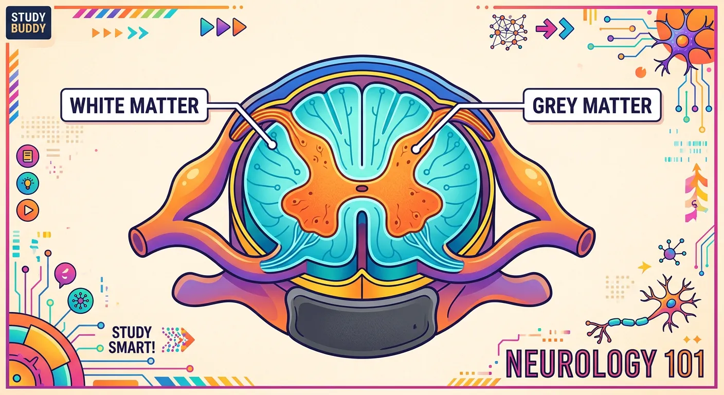
Within the subarachnoid space flows the Cerebrospinal Fluid (CSF). This fluid is more than just a cushion; it acts as a mechanical shock absorber, helps in nutrient distribution, and removes metabolic waste products from the delicate neural tissue. If you look at a cross-section of the spinal cord, you will see a distinct arrangement that is the opposite of the brain: 'grey matter' (cell bodies) forms an H-shaped inner core, while 'white matter' (myelinated axons) makes up the outer layers. The central canal, located in the middle of the grey matter, is also filled with CSF.
This structural organization allows the spinal cord to process immediate sensory inputs via the dorsal root and relay them to the brain, while simultaneously conveying motor commands via the ventral root back down to the body's limbs. Without the spinal cord's rapid relay capabilities, the brain would be functionally isolated, rendering us unable to move, sense environment changes, or survive. Mastering the order of the protective layers and the distribution of matter within the cord is essential for understanding clinical conditions like meningitis or spinal cord injuries.
Quick Revision Points
- Protective Layers: Dura mater (outer), Arachnoid (middle), Pia mater (inner).
- CSF: Located in the subarachnoid space; provides buoyancy and protection.
- Gray Matter: Inner H-shaped core containing nerve cell bodies and interneurons.
- White Matter: Outer area containing myelinated ascending and descending tracts.
- Roots: Dorsal root (sensory/afferent) and Ventral root (motor/efferent).
NEET Exam Angle
- Always remember the order of meninges from outer to inner (Dura -> Arachnoid -> Pia) using the mnemonic 'DAP'.
- Understand the matter arrangement: In the brain, gray is outer; in the spinal cord, gray is inner.
| Protective Component | Primary Function |
|---|---|
| Vertebral Column | Mechanical protection and skeletal support |
| Meninges (3 layers) | Biological containment and infection barrier |
| CSF | Cushioning, buoyancy, and nutrient exchange |
| Central Canal | Circulation of CSF within the spinal cord |
06Reflex Action and the Reflex Arc: Rapid Involuntary Responses
“Ever pulled your hand away from a hot stove before even thinking about it? That’s a reflex arc! The signal hits the spinal cord, and it sends a direct command to your muscles. It skips the long brain-processing step to keep you safe instantly!”
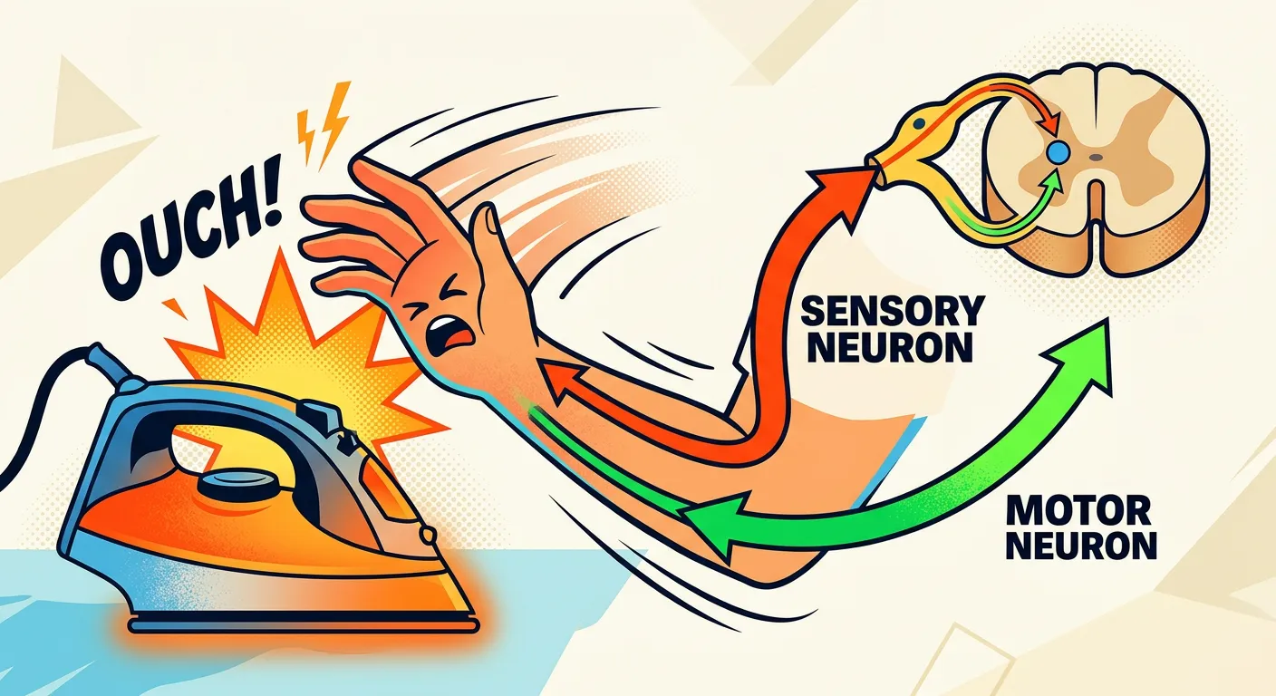
Reflex actions are the nervous system's way of bypassing 'central bureaucracy' for the sake of speed. When you touch a hot stove or step on a sharp object, you don't wait for your brain to deliberate on the danger; your body reacts instantly. This happens through the reflex arc—a specialized neuronal pathway that processes the stimulus at the level of the spinal cord without conscious effort. It consists of five essential components: a receptor that senses the stimulus, a sensory (afferent) neuron, an integration center (synapse in the spinal cord), a motor (efferent) neuron, and an effector (usually a muscle) that performs the action.
By skipping the long trip to the cerebral cortex for processing, the reflex arc saves precious milliseconds, which can be the difference between a minor burn and severe, permanent tissue damage. This is a classic example of an involuntary action. Once the spinal cord triggers the withdrawal, the sensory information is then relayed to the brain, which is why you feel the pain of the heat just a fraction of a second after you have already pulled your hand away. This delay proves the spinal cord acted independently of conscious thought.
Understanding the reflex arc is vital for NEET because it demonstrates the efficiency of localized neural control. Common examples include the knee-jerk reflex, the pupil's contraction in bright light, and the withdrawal reflex from thermal stimuli. These arcs are hardwired survival mechanisms that function even when the conscious brain is focused on other tasks. You should be able to trace the flow of an impulse from the sensory receptor through the dorsal root ganglion into the spinal cord, and out through the ventral root to the effector muscle.
Quick Revision Points
- Reflex Arc: Receptor -> Sensory Neuron -> Spinal Cord (Interneuron) -> Motor Neuron -> Effector.
- Function: Provides rapid, involuntary protection against immediate physical threats.
- Key Examples: Knee-jerk reflex (monosynaptic), withdrawal reflex (polysynaptic).
- Bypass logic: Direct spinal response allows for reactions faster than conscious perception.
NEET Exam Angle
- Be able to sequence the components of the reflex arc in the correct order of signal flow.
- Identify the 'Dorsal Root Ganglion' as the location of the cell bodies of sensory neurons.
- Recognize that the brain is notified of the event only after the reflex action has initiated.
| Component | Role in Reflex Arc |
|---|---|
| Receptor | Detects stimulus (e.g., heat, pressure, pain) |
| Sensory Neuron | Carries impulse via dorsal root to spinal cord |
| Integration Center | Synapse within the gray matter of the spinal cord |
| Motor Neuron | Carries command via ventral root to the muscle |
| Effector | The muscle or gland that executes the response |
07Neural Control and Integration: Critical Concepts for NEET Preparation

“And that is the magic of the nervous system! From processing complex NEET physics problems to feeling the excitement of a win, it is your greatest asset. Keep revising these basics, stay curious, and you will absolutely ace your biology goals. See you in the next lesson!”
As we conclude our exploration of the nervous system, it is important to reflect on how this system serves as the ultimate orchestrator of human physiology. It maintains homeostasis by integrating diverse sensory inputs with precise motor outputs, allowing for complex activities ranging from simple survival to abstract problem-solving and memory retrieval. For a NEET aspirant, the key to success lies in connecting these neural structures with their functional outcomes. For instance, you should understand how the hypothalamus links the nervous system to the endocrine system, or how the medulla oblongata works in concert with the lungs and heart to regulate your internal environment.
In your preparation, practice drawing diagrams of the neuron and the brain repeatedly. Labeling parts like the Nissl’s granules, the Nodes of Ranvier, and the specific lobes of the brain will help solidify your understanding. The more you visualize the pathway of an impulse—from the dendrites of one neuron, across the synaptic cleft, and through the spinal cord—the better you will retain these concepts under exam pressure. Don't let the technical terminology like 'repolarization' or 'corpora quadrigemina' overwhelm you; break it down into the core themes of 'command center' (CNS) and 'messengers' (PNS).
Always relate what you learn to real-world examples. Think of the reflex arc when you pull your hand from heat, or the role of the cerebellum when you see a gymnast balancing on a beam. NEET is less about rote memorization and more about understanding integrated systems. If you can explain why white matter is external in the spinal cord but internal in the brain, or how the Na+/K+ pump maintains a resting potential, you are thinking like a doctor. Keep revising the high-yield areas we discussed—especially the hypothalamus, synapse mechanisms, and neuron types. Stay consistent, use flowcharts for the reflex arc, and remember that mastering the neural system is a major milestone in your path to a medical career. Every hour spent understanding these biological circuits brings you one step closer to acing the exam.
Quick Revision Points
- Integration: The brain combines multiple sensory inputs for a single coordinated response.
- Homeostasis: Maintained via feedback loops involving the hypothalamus and autonomic system.
- Visualizing structure: Use labeled diagrams for brain regions and the reflex arc pathway.
- Matter Distribution: Brain (Gray-Outer, White-Inner); Spinal Cord (Gray-Inner, White-Outer).
- Consistency: Focus on recurring themes: Synapse, Medulla, and Hypothalamus functions.
NEET Exam Angle
- Focus on 'link' questions that connect nervous system structure to muscular or hormonal output.
- Prioritize diagrams, as labeled identification of neural structures is a consistent pattern in NEET papers.
- Understand the role of the Limbic system in memory and emotion, as this is a common high-yield topic.
| Study Habit | Benefit for NEET |
|---|---|
| Drawing Diagrams | Better retention of structural details and labels |
| Flowcharting | Clarity on complex impulse pathways and reflex arcs |
| PYQ Analysis | Identifying recurring themes like Hypothalamus and Synapse |
| Comparative Tables | Clearing confusion between CNS/PNS and matter distribution |
Recommended Reading
Explore related Biology topics to build deeper chapter connections for NEET.
- Morphology and Modifications · Topic 2.1
- Families · Topic 2.10
- Animal Tissues · Topic 2.11
- Frog Morphology · Topic 2.12
- Digestive System · Topic 2.13
- Circulatory System · Topic 2.14
- Jump to Key Terms (Quick Revision)
- Review Common NEET Mistakes
- Read Topic FAQs
- Check PYQ Pattern Notes
- Practice NEET MCQs
- Solve NEET PYQs
📚 Key Terms
⚠️ Common NEET Mistakes
- 1Confusing the location of gray and white matter: remember gray is inner in the spinal cord, but outer in the brain.
- 2Assuming neurotransmitters are involved in electrical synapses; they are only used in chemical synapses.
- 3Misremembering the order of the three meningeal layers; think 'DAP' for Dura, Arachnoid, Pia (outer to inner).
- 4Believing that reflexes involve the brain during the initial withdrawal response; the spinal cord handles the immediate action.
📝 NEET PYQ Pattern
In NEET 2018–2024, questions frequently focus on the functions of the Hypothalamus and the role of the Limbic system in emotions. Reflex arc diagrams and the location of specific neuron types (like bipolar neurons in the retina) are recurring high-yield themes that appear nearly every year.
❓ Frequently Asked Questions
What is the difference between an electrical and a chemical synapse?
Electrical synapses involve gap junctions allowing ions to flow directly between neurons for near-instant transmission, while chemical synapses rely on neurotransmitters (like acetylcholine) to bridge the synaptic cleft, allowing for more regulated, unidirectional signaling.
Which part of the brain is responsible for maintaining body temperature and hunger?
The hypothalamus, located in the forebrain, is the primary control center for homeostasis, including the regulation of body temperature, hunger, thirst, and various endocrine functions.
How does the structure of a bipolar neuron differ from a multipolar neuron, and where is it found?
A bipolar neuron has one axon and one dendrite, typically found in the retina of the eye, whereas a multipolar neuron has one axon and many dendrites, typically found in the cerebral cortex.
What is the function of the Medulla Oblongata in the hindbrain?
The medulla oblongata acts as the control center for vital involuntary life functions, including respiration, cardiovascular regulation, and gastric secretions.
Explain the sequence of components in a typical reflex arc path.
A reflex arc follows this sequence: Receptor detects the stimulus -> Sensory neuron carries the impulse -> Integration center (in spinal cord) processes it -> Motor neuron transmits the command -> Effector (muscle) executes the response.
Why is the myelination of neurons significant for nerve impulse transmission?
Myelin acts as an electrical insulator, allowing the nerve impulse to 'jump' between the Nodes of Ranvier in a process called saltatory conduction, which significantly increases the speed of transmission.
Written By
NEET Content Strategist & Biology Expert
Sangita Kumari is a NEET educator and content strategist with over 6 years of experience teaching Biology, Chemistry, and Physics to Class 11 and 12 aspirants. She helps bridge the gap between traditional NCERT preparation and modern AI-powered learning. Her content is trusted by thousands of NEET aspirants across India.