🎬 Video Lesson Available
Watch the full 7-slide video lesson for Reproductive System with AI teacher narration and visual explanations.
01The Biological Blueprint: Introduction to Human Reproductive Physiology
“Welcome, future doctors! Reproduction is the miracle of life, ensuring our species continues. Think of the reproductive system as the ultimate biological factory, meticulously designed to create the next generation. Today, we will decode this complex machinery, focusing on the structures essential for NEET success.”
Welcome to one of the most vital chapters in your NEET preparation. When we talk about the Reproductive System, we aren't just discussing a set of organs; we are looking at the 'Biological Blueprint' for the continuity of life. In the context of Class 11 Biology, understanding the structural organization of these systems is the first step toward mastering the complex hormonal and physiological processes you will encounter in Class 12. Reproduction is the fundamental mechanism that ensures a species does not go extinct, allowing for the transfer of genetic blueprints from one generation to the next. It is a process that balances the preservation of the species with the introduction of genetic variation through sexual reproduction.
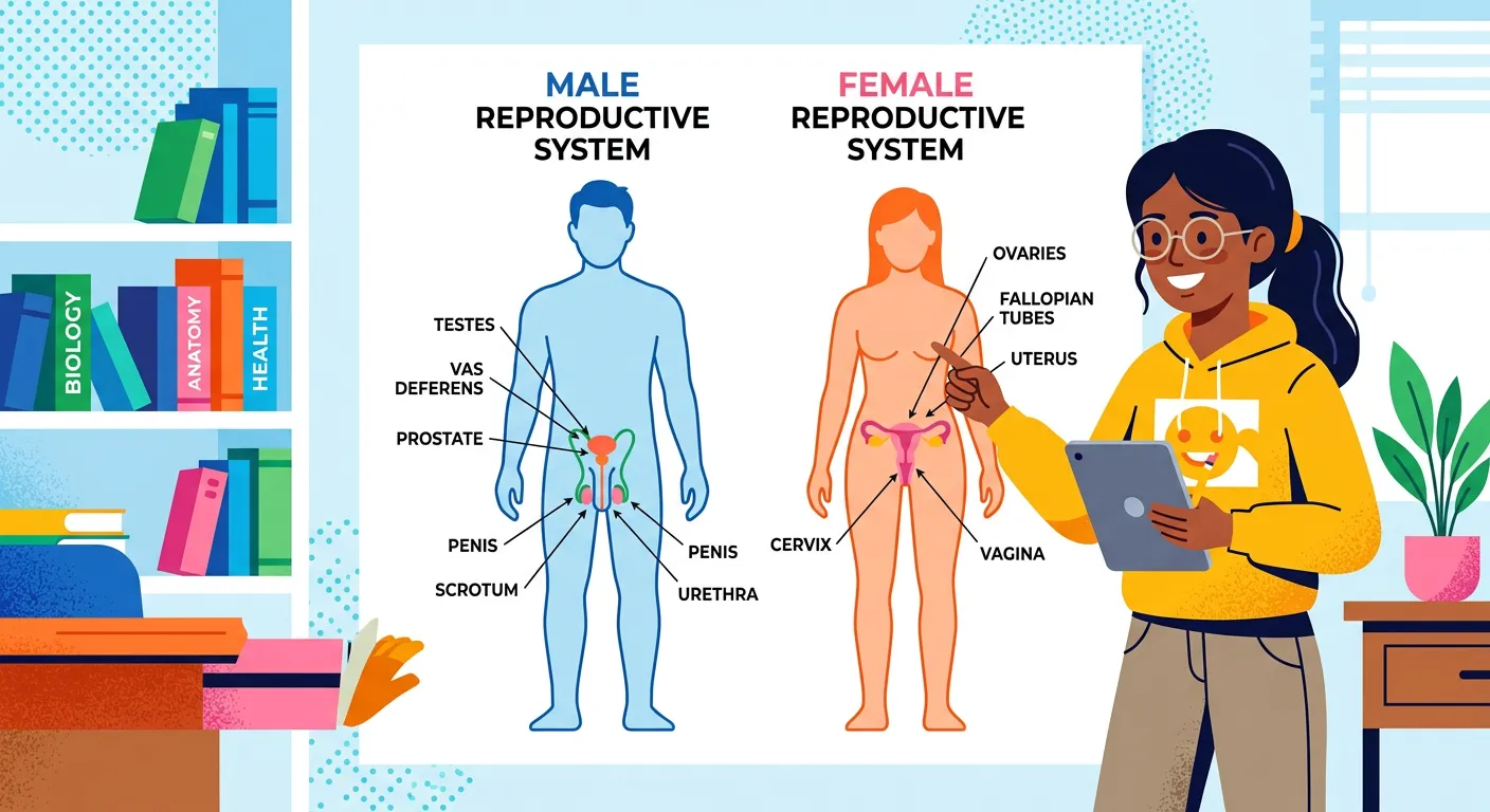
Think of the human reproductive system as a high-precision biological factory. It is designed with incredible specificity, involving specialized cells (gametes), complex duct systems for transport, and glandular tissues that regulate the entire process. For your exams, it is crucial to distinguish between primary and secondary sexual organs. Primary organs, like the testes and ovaries, are where the 'magic' of gamete production happens. Secondary organs, such as the fallopian tubes or the vas deferens, serve as the infrastructure—the highways and storage units that facilitate the meeting and maturation of these gametes. Without this infrastructure, the gametes would have no way to fulfill their biological destiny.
From an evolutionary standpoint, mammalian reproduction has evolved to be highly efficient. Features like internal fertilization and specialized structures for nourishing the developing embryo (viviparity) have given humans and other mammals a significant survival advantage. This efficiency is maintained by a sophisticated feedback loop between the brain and the gonads. As future medical professionals, you must view these structures not just as static labels on a diagram, but as a coordinated, dynamic system working in perfect harmony to ensure that life persists across millennia.
Quick Revision Points
- Reproduction ensures species continuity and introduces genetic variation.
- Primary sex organs (Gonads) produce gametes and secrete essential sex hormones.
- Secondary sex organs are essential for the transport, storage, and maturation of gametes.
- Human reproduction involves internal fertilization and the birth of live young (viviparity).
- The system is under strict endocrine control, primarily from the hypothalamus, pituitary, and gonads.
NEET Exam Angle
- Conceptual Focus: Differentiating between primary and secondary organs is a frequent 'Statement-based' question in the NEET biology section.
- High-Yield Fact: Remember that the gonads (testes/ovaries) have a dual function: cytogenic (producing gametic cells) and endocrine (producing hormones).
| Organ Category | Examples in Males | Examples in Females |
|---|---|---|
| Primary Sex Organs | Testes | Ovaries |
| Secondary Sex Organs | Vas deferens, Seminal vesicles | Fallopian tubes, Uterus |
| External Genitalia | Penis, Scrotum | Vulva |
02Male Reproductive Anatomy: The Testes and Thermoregulation Mechanics
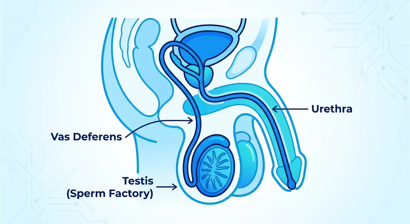
“Meet the male reproductive system. The star of the show is the Testis, our 'Sperm Factory'. Located outside the body in the scrotum, it keeps temperature lower than the body—crucial for healthy sperm production. It is not just about cells; it is about biological precision.”
In the male reproductive system, the focal point is undoubtedly the testes. These are paired, oval-shaped organs situated outside the abdominal cavity within a sac-like structure called the scrotum. Each testis measures about 4 to 5 cm in length and 2 to 3 cm in width. Why are they located outside the body? This is a classic NEET question. The process of sperm production, or spermatogenesis, is extremely sensitive to heat. Our core body temperature (37°C) is actually too high for sperm to develop properly. The scrotum acts as a thermoregulator, maintaining the testes at a temperature 2–2.5°C lower than the internal body temperature, which is the 'Goldilocks zone' for healthy sperm.
Sometimes, during fetal development, the testes fail to descend from the abdomen into the scrotum. This clinical condition is known as cryptorchidism. If left untreated, it can lead to sterility because the high internal body temperature prevents sperm maturation and can eventually lead to testicular cancer. This highlights the scrotum's critical biological role beyond just being a protective pouch; it is a thermal regulatory environment essential for male fertility. This anatomical placement is a hallmark of most mammals and represents a specialized adaptation for reproductive success.
Structurally, each testis is covered by a dense fibrous coating called the tunica albuginea and is divided into about 250 compartments called testicular lobules. Within these lobules, the real work happens inside one to three highly coiled seminiferous tubules. From there, the sperm travel through a specific pathway: the Rete testis, then the Vasa efferentia, followed by the Epididymis (where they mature and gain motility), and finally the Vas deferens. Mastering this anatomical sequence—from production to delivery—is a non-negotiable for anyone aiming for a top NEET score.
Quick Revision Points
- Testes are located in the scrotum for thermoregulation (2-2.5°C below core temp).
- Cryptorchidism is the failure of testes to descend into the scrotum, leading to potential sterility.
- The testes serve a dual role: producing sperm and secreting the androgen testosterone.
- The Epididymis is the primary site for sperm maturation and temporary storage.
- The duct system pathway: Seminiferous tubules → Rete testis → Vasa efferentia → Epididymis → Vas deferens.
NEET Exam Angle
- Pattern Alert: Chronological 'flowchart' questions on the sperm pathway appear almost every alternate year. Ensure you don't skip the Vasa efferentia.
- Clinical Relevance: Questions often link the temperature of the scrotum to male infertility or the clinical implications of cryptorchidism.
03Microscopic Architecture: Seminiferous Tubules and Spermatogenesis
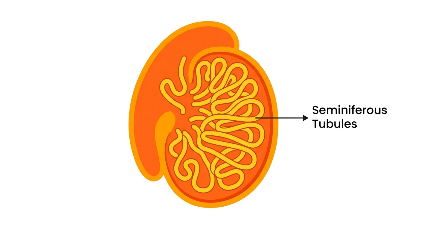
“Inside the testes, we find coiled structures called seminiferous tubules. Think of these as the assembly lines where spermatogenesis happens. Millions of sperm are produced here daily. It is a highly organized, non-stop production process, vital for carrying genetic blueprints to the next generation.”
When we zoom into the microscopic level of the testes, we find the seminiferous tubules—the actual 'assembly lines' of sperm production. The wall of each tubule is lined with two distinct types of cells: the male germ cells (spermatogonia) and the Sertoli cells. The spermatogonia undergo meiotic divisions to eventually transform into mature spermatozoa. However, these developing cells are fragile and need constant support. This is where the Sertoli cells, often called 'nurse cells,' come into play. They provide the necessary nutrition, structural support, and paracrine signaling to the germ cells during their transformation. They also form the blood-testis barrier, protecting the developing sperm from the immune system.
Outside the tubules, in the narrow interstitial spaces, we find another hero of male physiology: the Leydig cells (or interstitial cells). These cells are responsible for synthesizing and secreting testicular hormones called androgens, the most famous being testosterone. This hormone is the fuel that drives spermatogenesis and maintains male secondary sexual characteristics like facial hair and voice deepening. The interaction between these cells is tightly regulated by hormones from the anterior pituitary—specifically LH, which acts on Leydig cells, and FSH, which acts on Sertoli cells.
Spermatogenesis is a continuous, non-stop process starting at puberty. Unlike females, who are born with a finite number of oocytes, males produce millions of sperm daily. This process requires a precise dance of cell division (mitosis for replenishment of the germ cell pool and meiosis for genetic reduction). Understanding the histology of the testes is essential for grasping how the body manages such a high-volume production of complex cells while maintaining genetic integrity. For NEET, being able to identify these cells on a histology slide is a key skill.
Quick Revision Points
- Seminiferous tubules are the functional units for sperm production and maturation.
- Sertoli cells provide nourishment and support to developing sperms (nurse cells).
- Leydig cells secrete testosterone in response to Luteinizing Hormone (LH).
- Spermatogenesis involves both mitosis (spermatogonia) and meiosis (spermatocytes).
- The interstitial space contains blood vessels, Leydig cells, and immune-competent cells.
NEET Exam Angle
- Common Trap: Students often confuse the location of Sertoli cells (inside the tubule) with Leydig cells (outside the tubule in the interstitial space).
- Visual ID: Be prepared to identify these cells on a cross-section diagram of a seminiferous tubule in the NCERT format.
| Cell Type | Location | Primary Function |
|---|---|---|
| Spermatogonia | Lining of Tubule | Undergo meiosis to form sperm |
| Sertoli Cells | Lining of Tubule | Support and nourish germ cells |
| Leydig Cells | Interstitial Space | Produce androgens (Testosterone) |
04Female Reproductive Essentials: Ovaries, Fallopian Tubes, and the Uterine Nest
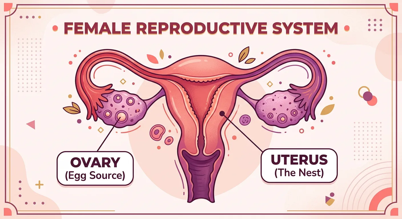
“Now, the female system. Here, the Ovaries act as the 'Egg Source', releasing one mature ovum periodically. The fallopian tubes are the meeting point, while the uterus is the 'Nest'—an incredibly resilient, muscular home designed to nourish and protect a developing baby for nine months.”
The female reproductive system is designed for a completely different set of challenges. While the male system is about mass production and delivery, the female system is focused on selection, nurturing, and protection. The ovaries are the primary sex organs, acting as the 'Egg Source.' Unlike the continuous production in males, oogenesis in females is cyclical and starts before birth, typically releasing just one mature ovum every month during the reproductive years. Each ovary is a solid organ covered by a thin epithelium-class-11-neet-biology)-class-11-neet-biology)-class-11-neet-biology)-class-11-neet-biology)-class-11-neet-biology)-class-11-neet-biology) and contains the ovarian stroma, divided into an outer cortex and an inner medulla.
Once released during ovulation, the ovum is picked up by the fimbriae—finger-like projections at the funnel-shaped edge (infundibulum) of the Fallopian tube (oviduct). The Fallopian tube is divided into three sections: the infundibulum, the wide ampulla, and the narrow isthmus. It's fascinating to note that the ovum is non-motile; it relies on the rhythmic beating of ciliated epithelium lining the tubes to move toward the uterus. This is a perfect example of how structural organization (animal tissues) directly supports physiological function. If the cilia are damaged, it can lead to complications like ectopic pregnancy.
Then we have the uterus, or the 'womb.' This is an incredibly resilient, pear-shaped organ composed of three distinct layers. The outermost is the thin perimetrium. The middle layer, the myometrium, is a thick layer of smooth muscle that generates the powerful contractions needed during childbirth (parturition). The innermost layer, the endometrium, is highly vascular and glandular. It is this layer that undergoes dramatic cyclical changes during the menstrual cycle and provides the 'nest' where an embryo can implant and grow into a fetus.
Quick Revision Points
- Ovaries produce the ovum and steroid hormones (Estrogen and Progesterone).
- Fimbriae help in the collection of the ovum from the peritoneal cavity after ovulation.
- Ciliated epithelium in the Fallopian tube facilitates the transport of the non-motile ovum.
- The Uterus consists of three layers: Perimetrium, Myometrium, and Endometrium.
- The Myometrium is responsible for strong labor contractions during delivery.
NEET Exam Angle
- High Probability: Questions regarding the layers of the uterus—specifically which one undergoes cyclical changes (Endometrium) vs. which one contracts (Myometrium).
- Pathway Focus: The correct sequence of the Fallopian tube parts: Infundibulum → Ampulla → Isthmus.
| Uterine Layer | Tissue Type | Function |
|---|---|---|
| Endometrium | Glandular/Vascular | Implantation and menstrual cycling |
| Myometrium | Smooth Muscle | Strong contractions during parturition |
| Perimetrium | Thin Membranous | Outer protective covering |
05The Spark of Life: Fertilization and Zygote Formation in the Ampulla
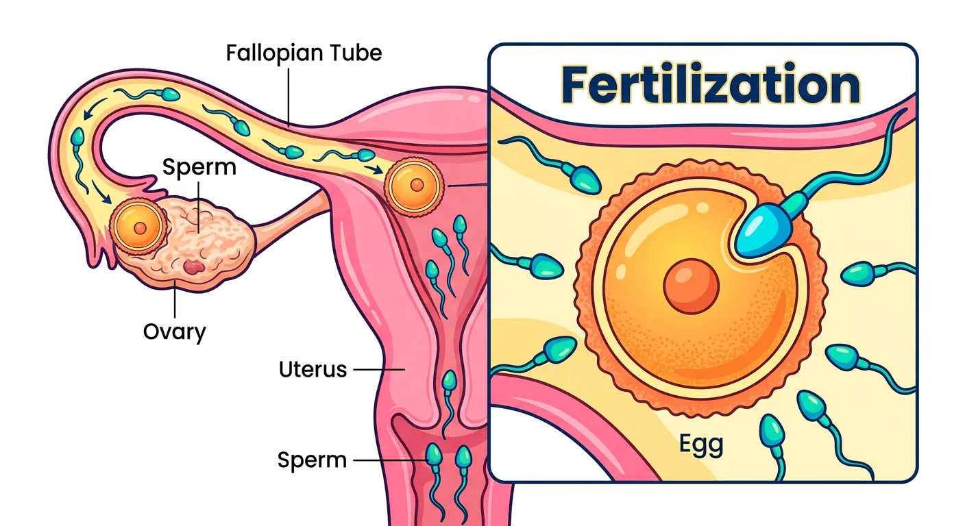
“The moment of truth: Fertilization! This usually happens in the ampulla of the fallopian tube. One lucky sperm fuses with the ovum to form a zygote. This union is the biological spark, combining genetic information from both parents to create a unique, brand-new individual.”
Fertilization is the pivotal moment where two separate genetic lineages combine to form a new individual. In humans, this 'spark of life' typically occurs in the ampulla of the Fallopian tube. You might often see older texts mentioning the 'ampullary-isthmic junction,' but the ampulla is recognized as the most accurate specific site for this event. For fertilization to be successful, both the sperm and the ovum must reach this junction simultaneously, which explains why not all acts of coitus lead to pregnancy.
It is not an easy journey for the sperm. They must first undergo 'capacitation' in the female tract—a process where they gain the ability to fertilize the egg. When a sperm contacts the egg, it undergoes the 'acrosome reaction,' releasing enzymes like hyaluronidase that digest the tough outer layers of the ovum (the corona radiata and the zona pellucida). The moment one sperm penetrates the zona pellucida, it triggers a 'block to polyspermy'—a rapid chemical change that prevents any other sperm from entering. This ensures that the resulting zygote is diploid (2n), with exactly 46 chromosomes, preventing lethal chromosomal abnormalities.
This process of syngamy (fusion of gametes) does more than just start a pregnancy; it triggers the completion of the second meiotic division in the ovum, which was previously arrested at the metaphase II stage. The resulting zygote is a single-celled powerhouse of potential, containing the unique genetic blueprint that defines everything from eye color to biochemical predispositions. It is the ultimate example of genetic recombination in action, creating a person who is genetically distinct from both parents.
Quick Revision Points
- The Ampulla is the primary site of human fertilization.
- Acrosomal enzymes (like Hyaluronidase) allow sperm to penetrate protective egg barriers.
- The Zona Pellucida layer prevents polyspermy through chemical modification (cortical reaction).
- Fertilization restores the diploid (2n) state from two haploid (n) gametes.
- Completion of the second meiotic division of the oocyte occurs only after sperm entry.
NEET Exam Angle
- Specific Location: If a question asks for the site of fertilization, 'Ampulla' is your top choice based on the latest NCERT interpretations.
- Process Detail: Be clear on the sequence: Sperm entry → Block to polyspermy → Completion of Meiosis II → Fusion of nuclei (Syngamy).
- Terminology: Understand 'capacitation'—the final maturation of sperm in the female tract required before fertilization can occur.
06Implantation Dynamics: Securing the Blastocyst in the Endometrial Wall
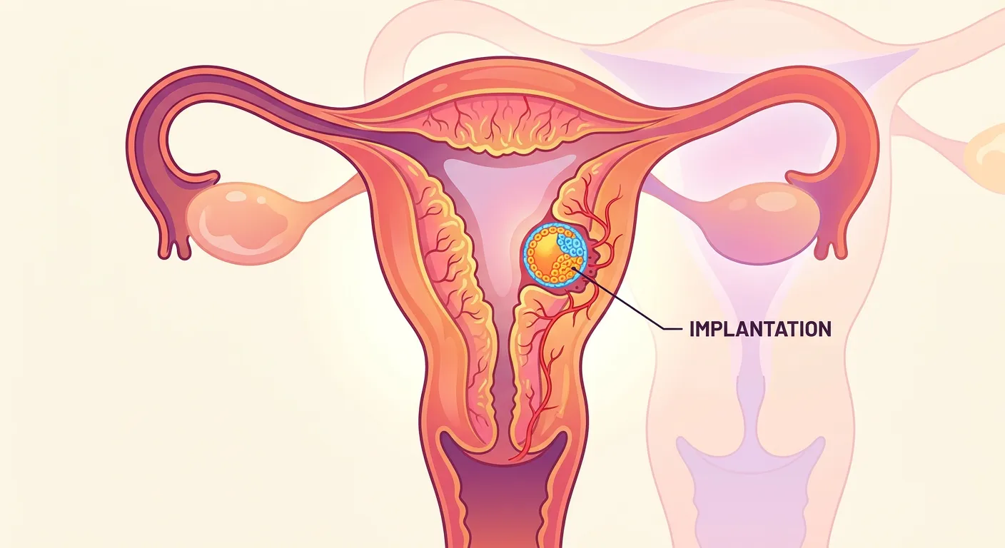
“After fertilization, the zygote travels to the uterus. It then undergoes 'Implantation,' embedding itself firmly into the nutrient-rich uterine wall, or endometrium. It is like planting a seed in fertile soil; from here, the incredible journey of fetal development truly begins. Growth is exponential!”
Once the zygote is formed, it doesn't just sit there. It immediately begins a journey toward the uterus, undergoing a series of rapid mitotic divisions called cleavage. Unlike regular mitosis, cleavage increases the cell number but the total size of the embryo remains almost the same. As it travels down the Fallopian tube, it transforms from a 2-cell stage to a 4-cell stage, then to an 8-cell stage (morula). Eventually, by the time it reaches the uterus, it becomes a hollow ball of cells known as the blastocyst. This is the stage that is ready for the next big step: implantation.
The blastocyst has a very specific structure that you must remember for NEET. It consists of an outer layer called the trophoblast and an inner cluster of cells called the inner cell mass (ICM). The trophoblast is what eventually forms the extra-embryonic membranes and the placenta by attaching to the endometrium. The inner cell mass is the 'source'—it contains the pluripotent stem cells that will differentiate into all the specialized tissues and organs of the embryo. This differentiation is the foundation of human development.
Implantation typically occurs about 7 days after fertilization. The blastocyst 'sinks' into the nutrient-rich, glandular endometrial wall, which then grows over it. For this to happen, the uterus must be receptive, a state maintained by the hormone progesterone (secreted by the corpus luteum in the ovary). If implantation is successful, the trophoblast begins to secrete HCG (Human Chorionic Gonadotropin), which signals the body to maintain the pregnancy. If you think of fertilization as the spark, implantation is the planting of the seed into fertile soil, initiating the long process of gestation.
Quick Revision Points
- Cleavage consists of rapid mitotic divisions without significant increase in total cytoplasmic volume.
- The Morula is a solid ball of 8-16 cells resembling a mulberry.
- The Blastocyst is the specific developmental stage that implants into the uterine wall.
- The Trophoblast layer helps in attachment and eventual placental formation.
- The Inner Cell Mass (ICM) gives rise to the actual embryo and its primary tissues.
NEET Exam Angle
- Structural ID: Questions often ask which part of the blastocyst becomes the embryo (Inner Cell Mass) and which part attaches to the mother (Trophoblast).
- Timing: Remember that implantation usually happens around Day 7 post-fertilization, after the morula stage.
| Stage | Cell Number / Description | Key Feature |
|---|---|---|
| Zygote | Single Cell | Diploid (2n) result of fusion |
| Morula | 8 to 16 cells | Solid mulberry-like ball |
| Blastocyst | ~100 cells (hollow) | Has Trophoblast and Inner Cell Mass |
| Implantation | N/A | Embedding into the Endometrium |
07NEET Strategy: Mastering the Sequence of Human Physiology
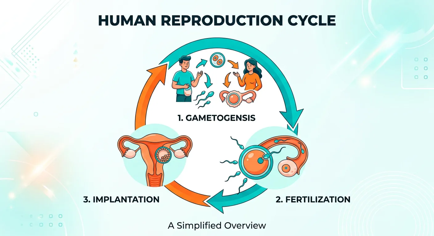
“To ace NEET, remember this sequence: Gametogenesis, Fertilization, and Implantation. Each step is a perfect biological relay race. Understanding this system is key to mastering human physiology. Keep revising these core concepts, and you will definitely shine in your exams. See you in the next lecture!”
As we wrap up this topic, let's focus on the 'big picture'—the strategy you need to tackle questions on the reproductive system. NEET loves sequences. You should be able to visualize the entire biological relay race without hesitation: Gametogenesis (forming the cells) → Insemination (transferring sperm) → Fertilization (fusion in the ampulla) → Zygote formation → Cleavage → Implantation (the uterine hug). This chronological flowchart is the backbone of almost all physiological questions in this unit. If you can track the path of a single cell from the testis to the uterus, you have mastered the fundamental anatomy required for the exam.
Another high-yield area is comparing male and female gamete production. Remember that male spermatogenesis is continuous and starts at puberty, whereas female oogenesis starts before birth, pauses during childhood, and restarts at puberty in a cyclical fashion. This distinction—the timing of meiosis—is often the basis for 'Statement A and Statement B' type questions. You must also distinguish between the primary hormones; while Testosterone drives the male system, the female system is a complex interplay between FSH, LH, Estrogen, and Progesterone. Understanding when these hormones peak is crucial for answering questions about the menstrual cycle, which you will study in more depth later.
Finally, pay close attention to the diagrams in your NCERT textbook. NEET examiners frequently take these diagrams and label them with 'A, B, C, D,' asking you to identify the parts or their specific functions. Focus specifically on the testis cross-section, the female reproductive tract, and the structure of the blastocyst. If you can draw these from memory and explain the role of each part, you are already ahead of 90% of the competition. Keep your revision consistent, and remember: biology is not about memorizing words, it's about understanding life's intricate machinery. Use flashcards for the hormonal peaks and the names of the accessory glands, as these are easy marks if you have them memorized. A common mistake is overlooking the secretory nature of the prostate and seminal vesicles, so ensure you review their specific chemical contributions to semen. By mastering these nuances, you ensure that no 'tricky' question can catch you off guard during the actual examination.
Quick Revision Points
- Correct Sequence: Gametogenesis → Insemination → Fertilization → Implantation.
- Sperm pathway: Seminiferous tubules → Rete testis → Epididymis → Vas deferens → Urethra.
- Hormonal control: Hypothalamus (GnRH) → Anterior Pituitary (FSH/LH) → Gonads.
- The corpus luteum is essential for early pregnancy maintenance through Progesterone secretion.
- Diagram labeling of the male and female systems is a guaranteed high-yield task for the NEET exam.
NEET Exam Angle
- High Frequency: Correct sequence questions regarding embryonic stages (Zygote → Morula → Blastocyst → Gastrula) are very common.
- Comparative Biology: Note the difference in the timing of meiosis completion between males (at maturation) and females (after fertilization).
- Last Minute Tip: Re-check the functions of the accessory glands (Seminal vesicles, Prostate, Bulbourethral glands)—they contribute to the 'semen' which protects and nourishes sperm.
Recommended Reading
Explore related Biology topics to build deeper chapter connections for NEET.
- Morphology and Modifications · Topic 2.1
- Families · Topic 2.10
- Animal Tissues · Topic 2.11
- Frog Morphology · Topic 2.12
- Digestive System · Topic 2.13
- Circulatory System · Topic 2.14
- Jump to Key Terms (Quick Revision)
- Review Common NEET Mistakes
- Read Topic FAQs
- Check PYQ Pattern Notes
- Practice NEET MCQs
- Solve NEET PYQs
📚 Key Terms
⚠️ Common NEET Mistakes
- 1Confusing Leydig cells (interstitial, produce testosterone) with Sertoli cells (intratubular, nourish sperm).
- 2Stating that fertilization occurs in the uterus instead of the Fallopian tube (ampulla).
- 3Thinking that the scrotum keeps the testes warmer than the body; it actually keeps them cooler.
- 4Incorrectly identifying the Morula as the stage that implants; it is the Blastocyst that implants.
- 5Forgetting that meiosis II in the ovum only completes after a sperm has entered the egg.
📝 NEET PYQ Pattern
Questions from 2018–2024 consistently target the specific site of fertilization (Ampulla) and the function of the scrotum in temperature regulation. There is a high frequency of 'Correct Sequence' questions regarding the pathway of sperm and embryonic development stages (Zygote → Morula → Blastocyst).
❓ Frequently Asked Questions
Why are the testes located outside the abdominal cavity in the scrotum?
The testes are located in the scrotum to maintain a temperature 2-2.5°C lower than the internal body temperature. This cooler environment is essential for the process of spermatogenesis (sperm production), as normal body temperature inhibits sperm development.
Where exactly does fertilization take place in the human female reproductive tract?
Fertilization typically occurs in the ampulla of the Fallopian tube (oviduct). It is the widest part of the tube where the sperm and ovum meet and fuse to form a zygote.
What is the difference between the myometrium and the endometrium of the uterus?
The endometrium is the innermost glandular layer that undergoes cyclical changes during the menstrual cycle and is the site of implantation. The myometrium is the middle thick layer of smooth muscle that produces strong contractions during the delivery of a baby.
What is the sequence of events from gamete production to implantation?
The correct sequence is: Gametogenesis (gamete production) → Insemination (transfer of sperm) → Fertilization (fusion of gametes in the ampulla) → Zygote formation → Cleavage (cell division) → Morula → Blastocyst → Implantation (embedding in the endometrium).
Which cells in the testes provide nourishment to the developing sperm?
Sertoli cells, also known as 'nurse cells,' are located inside the seminiferous tubules and provide structural support and nourishment to the developing male germ cells (spermatogonia) during spermatogenesis.
What defines the process of implantation and when does it occur?
Implantation is the process where the blastocyst (at the 7th-day stage) embeds itself into the endometrial lining of the uterus. It usually occurs about 7 days after fertilization and is a prerequisite for a successful pregnancy.
Written By
NEET Content Strategist & Biology Expert
Sangita Kumari is a NEET educator and content strategist with over 6 years of experience teaching Biology, Chemistry, and Physics to Class 11 and 12 aspirants. She helps bridge the gap between traditional NCERT preparation and modern AI-powered learning. Her content is trusted by thousands of NEET aspirants across India.