🎬 Video Lesson Available
Watch the full 7-slide video lesson for Tissues with AI teacher narration and visual explanations.
01Defining the Biological Foundation: Cellular Cooperation and Tissue Formation
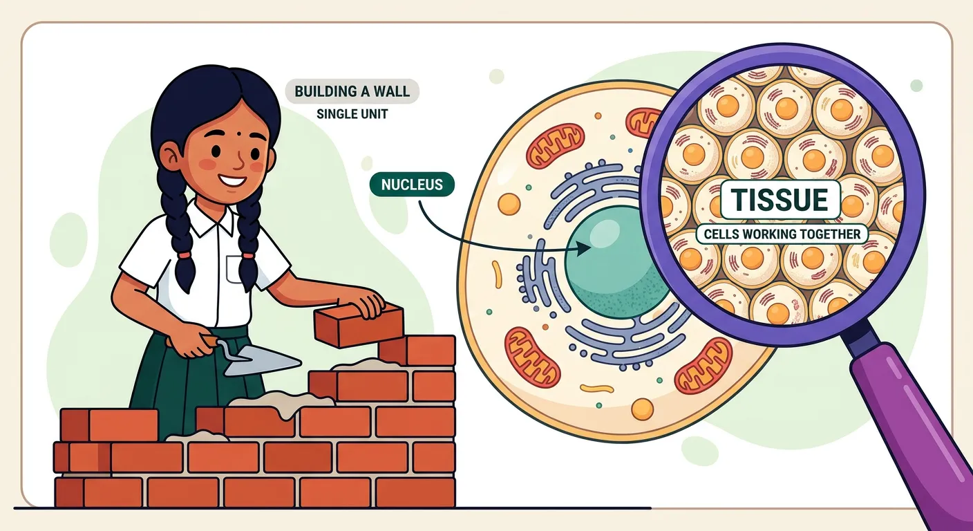
“Welcome! Imagine building a house. One brick is a cell, but the whole wall is a tissue. In biology, a tissue is a group of similar cells working together to perform a specific function. Like players in a cricket team, they unite to win the match!”
When we look at the complexity of a multicellular organism, it is easy to get overwhelmed by the sheer number of processes happening simultaneously. However, biology operates on a beautifully organized hierarchy-and-systematics). It starts with the cell, the basic unit of life. But a single cell, no matter how specialized, cannot sustain the life of a complex animal like a human or a lion. This is where the concept of a 'tissue' becomes critical. A tissue is essentially a group of similar cells, along with their intercellular substances, which originate from the same embryonic layer and work together to perform a specific, coordinated function.
In the evolutionary timeline, the transition from unicellular to multicellular life required a 'division of labor.' Think of it like a cricket team: one player cannot be the bowler, the wicketkeeper, and the entire batting lineup all at once. Instead, individuals specialize in roles to ensure the team wins. Similarly, in our bodies, cells cluster into tissues to handle specific tasks like movement, sensation, or protection. This organization allows for greater efficiency and the development of complex organs. When you study this for NEET, remember that the interaction between these cells is just as important as the cells themselves.
Understanding tissues is the gateway to mastering human physiology. If you know how the 'bricks' (cells) and the 'mortar' (intercellular matrix) are arranged to form a 'wall' (tissue), you will intuitively understand why a lung can exchange gases while a bicep can only pull a bone. This section serves as your foundation for Chapter 8 (Cell: The Unit of Life) and prepares you for the structural complexity seen in later chapters.
Quick Revision Points
- Tissue Definition: A group of similar cells and intercellular substances performing a specific function.
- Cellular Hierarchy: Cells → Tissues → Organs → Organ Systems → Organism.
- Division of Labor: Essential for multicellular organisms to maintain homeostasis and efficiency.
- Common Origin: Cells within a tissue typically share the same embryonic germ layer (Ectoderm, Mesoderm, or Endoderm).
- Intercellular Matrix: The non-living material between cells that provides structural and biochemical support.
NEET Exam Angle
- Conceptual Clarity: NEET often tests the fundamental definition of a tissue. Remember, similarity in function is usually paired with a common origin.
- Evolutionary Context: Questions may arise regarding why multicellularity requires tissue specialization (Efficiency and Division of Labor).
- Linkage: Be prepared to link cellular organelles (like mitochondria) to tissues that require high energy (like muscle tissue).
02The Four Pillars of Animal Architecture: Classification and Primary Functions
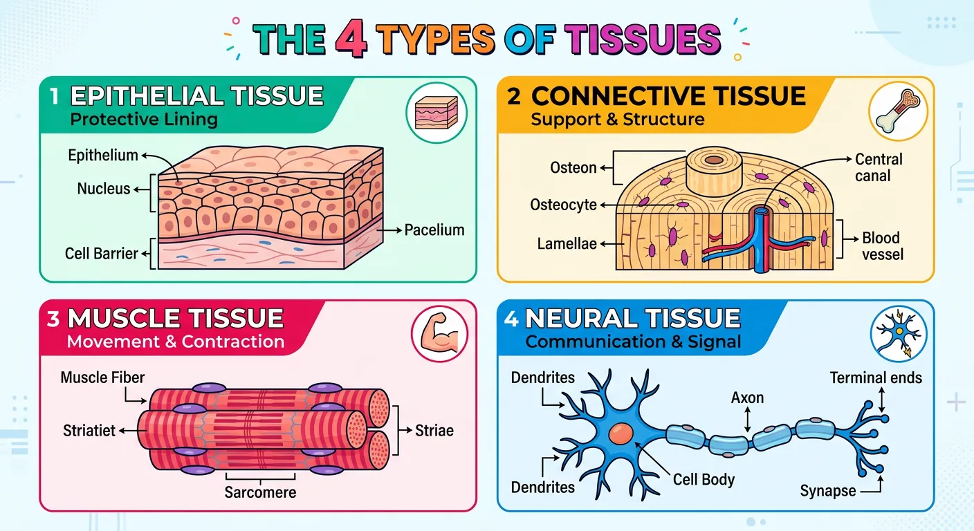
“In the human body, we have four main tissue types: Epithelial, which covers us like skin; Connective, the glue holding everything together; Muscle, for movement; and Neural, our body's communication network. Think of these as the four pillars supporting our complex biological architecture.”
The animal kingdom exhibits a vast diversity of forms, yet every single complex animal is built using only four basic types of tissues. These are the Epithelial, Connective, Muscular, and Neural tissues. You can think of these as the 'four pillars' of animal architecture. Each type is uniquely designed to serve a distinct physiological need. Epithelial tissue acts as the covering or lining; Connective tissue provides support and binds structures; Muscular tissue enables movement through contraction; and Neural tissue facilitates communication by transmitting electrical impulses.
From a NEET perspective, understanding the embryonic origin of these tissues is paramount. While most tissues derive from a specific germ layer, some are more diverse. For example, epithelial tissue is unique because it can originate from all three germ layers: Ectoderm (skin epidermis), Mesoderm (lining of coelom), and Endoderm (lining of the gut). Connective and Muscular tissues are primarily Mesodermal in origin, whereas Neural tissue is almost exclusively Ectodermal. Mastering this 'origin map' helps you predict the behavior and location of these tissues throughout the body.
These four tissues do not work in isolation. They are intricately interconnected to maintain homeostasis. For instance, the nervous system (Neural) sends a signal to a bicep (Muscle), which is supported by a bone (Connective), all while being protected by the skin (Epithelial). This integration is what makes life possible. When you revise this, focus on how the structure of each tissue type is a direct reflection of its primary function.
| Tissue Type | Primary Function | Embryonic Origin |
|---|---|---|
| Epithelial | Protection, Secretion, Absorption | Ectoderm, Mesoderm, Endoderm |
| Connective | Support, Binding, Transport | Mesoderm |
| Muscular | Movement and Locomotion | Mesoderm |
| Neural | Control and Coordination | Ectoderm |
Quick Revision Points
- Classification: Four types: Epithelial, Connective, Muscular, and Neural.
- Epithelial Origin: The only tissue type that arises from all three germ layers.
- Connective Dominance: Connective tissue is the most abundant and widely distributed in the body.
- Neural Specialization: Specialized for excitability and conductivity to manage rapid responses.
- Homeostasis: The collective action of these tissues keeps the internal environment stable.
NEET Exam Angle
- Embryonic Origins: Frequent MCQs target the origin of specific tissues (e.g., 'Which tissue is mesodermal in origin?').
- Identification: Be ready to classify a described function (like 'binding of organs') into one of the four main categories.
- Integration Questions: Questions often link these tissues to the Human Physiology unit, requiring a 'big picture' understanding.
03Epithelial Tissue: Structural Variants and Protective Gatekeepers
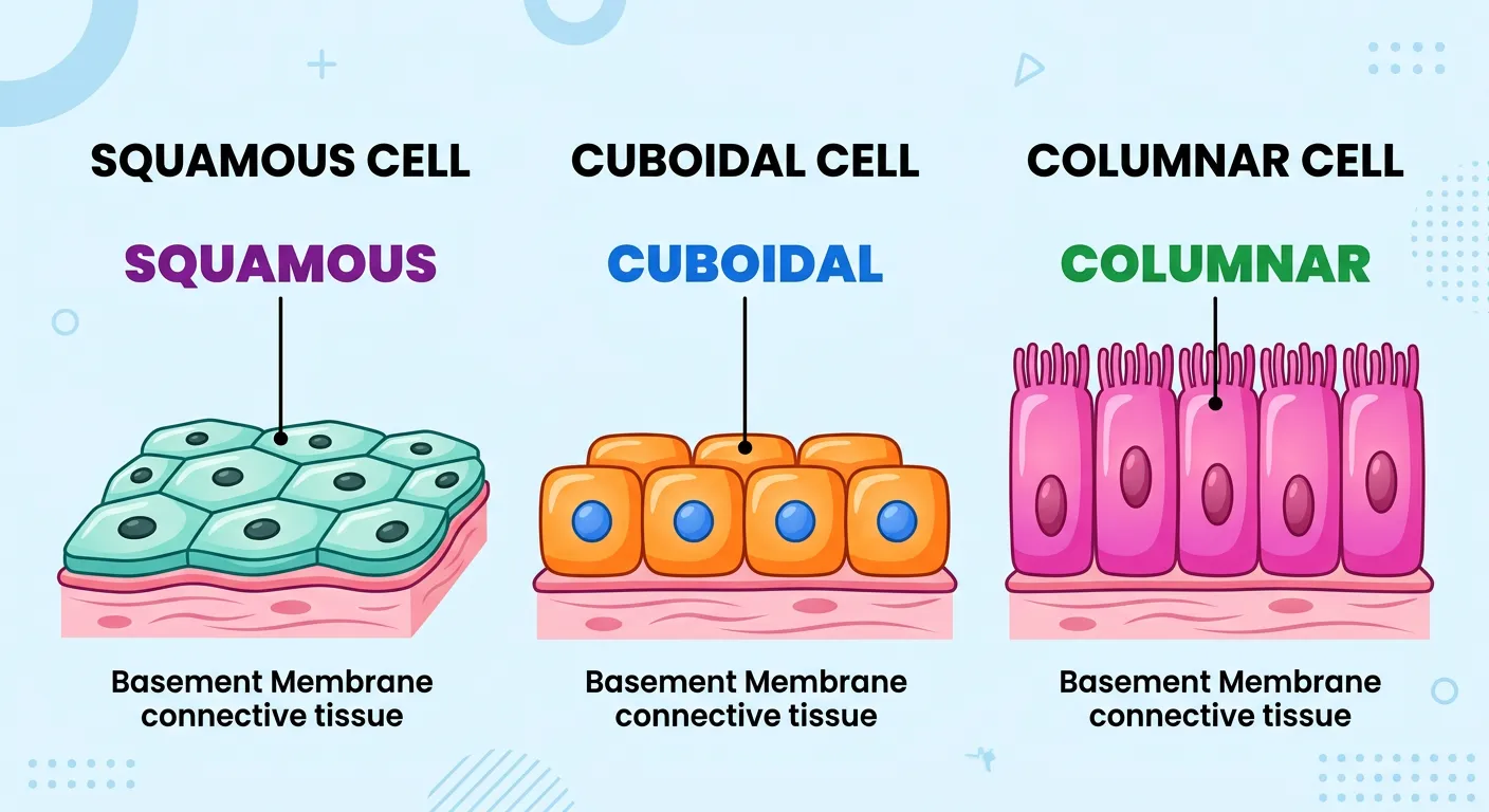
“Epithelial tissue is our protective shield! Squamous are flat like floor tiles, Cuboidal are boxy like dice, and Columnar are tall like pillars. They line our organs and skin, acting as the ultimate gatekeepers, deciding exactly what enters or leaves our body's internal systems.”
Epithelial tissue, or epithelium, is the body's primary interface with the environment. Whether it's the outer surface of your skin or the inner lining of your stomach, epithelium is always there. One of its defining characteristics is that the cells are compactly packed with very little intercellular matrix. This 'wall-like' structure makes it an excellent protective barrier. Every epithelial layer rests on a non-cellular 'basement membrane,' which provides structural support and anchors it to the underlying connective tissue.
We classify epithelial tissues based on the number of layers and the shape of the cells. 'Simple epithelium' consists of a single layer and is typically found where absorption or filtration occurs. 'Compound (Stratified) epithelium' has multiple layers and serves a protective role, like in our skin. Within these categories, we look at cell shapes: Squamous (flat), Cuboidal (cube-like), and Columnar (tall). Each shape is a functional adaptation. Squamous cells allow for easy diffusion (like in the lungs), while Cuboidal and Columnar cells often feature microvilli to increase surface area for absorption in the gut or kidneys.
Another critical feature for NEET is the presence of cell junctions. Tight junctions prevent leakage, Adhering junctions perform 'cementing' to keep cells together, and Gap junctions allow for rapid communication between adjacent cells. These junctions ensure that the epithelium functions as a cohesive unit rather than just a collection of independent cells.
| Epithelial Type | Structural Feature | Common Location | Primary Function |
|---|---|---|---|
| Simple Squamous | Flat, scale-like cells | Blood vessels, Alveoli | Diffusion & Filtration |
| Simple Cuboidal | Cube-shaped cells | Kidney tubules (PCT) | Secretion & Absorption |
| Simple Columnar | Tall, pillar-like cells | Stomach, Intestine | Absorption & Secretion |
| Ciliated | Presence of Cilia | Bronchioles, Fallopian tubes | Moving particles/mucus |
Quick Revision Points
- Simple vs. Compound: Single layer vs. multiple layers.
- Basement Membrane: A thin, fibrous, non-cellular matrix that anchors the epithelium.
- Cell Junctions: Tight (prevent leak), Adhering (anchor), Gap (communicate).
- Avascularity: Epithelial tissue lacks blood vessels; it gets nutrients via diffusion from underlying connective tissue.
- Microvilli: Finger-like projections on cuboidal/columnar cells to increase absorption efficiency.
NEET Exam Angle
- Location-Based Questions: 'Where is ciliated epithelium found?' is a classic NEET question (Ans: Bronchioles/Fallopian tubes).
- Diagram Identification: Identify squamous, cuboidal, or columnar cells from NCERT-based images.
- Junction Functions: Match the junction type (Gap, Tight, Adhering) to its specific physiological role.
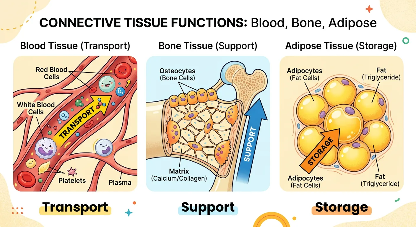
04Connective Tissue: The Abundant Glue and Structural Framework
“Connective tissue is the most abundant type! From the hard bones supporting our frame to blood, the liquid highway transporting oxygen, and adipose tissue storing fat like a reserve fuel tank. It’s the 'connective glue' that keeps our entire body structure organized and functional.”
Connective tissue is the most abundant and widely distributed tissue in the body of complex animals. Its primary role is exactly what the name suggests: it 'connects' and supports other tissues and organs. Unlike epithelial tissue, connective tissue has a massive amount of extracellular matrix, within which various cells and fibers are suspended. This matrix consists of ground substance (modified polysaccharides) and protein fibers like Collagen (for strength) and Elastin (for flexibility).
Connective tissues are categorized into three main types: Loose, Dense, and Specialized. Loose connective tissues, like Areolar and Adipose, have fibers and cells loosely arranged. Areolar tissue acts as a support framework for epithelium, while Adipose tissue is a specialized storage site for fats. Dense connective tissues, such as Tendons (muscle to bone) and Ligaments (bone to bone), are packed with collagen fibers to withstand high tension. Finally, Specialized connective tissues include Bone, Cartilage, and Blood. Bone and Cartilage provide the skeletal framework, while Blood is a unique fluid tissue that lacks fibers in its normal state and functions as the body's transport system.
For NEET aspirants, it is vital to distinguish between 'white fibrous' (collagen-rich) and 'yellow elastic' (elastin-rich) tissues. Collagen provides the tensile strength needed for tendons, while elastin allows the skin and arteries to snap back into place after being stretched. Remember, all connective tissues except blood secrete structural proteins (fibers).
Quick Revision Points
- Matrix Components: Ground substance + Fibers (Collagen/Elastin) + Cells (Fibroblasts, Macrophages, Mast cells).
- Areolar Tissue: Found beneath the skin; serves as a reservoir of water and salts.
- Adipose Tissue: Specializes in fat storage; provides insulation and shock absorption.
- Tendons vs. Ligaments: Tendons connect Muscle to Bone; Ligaments connect Bone to Bone.
- Specialized Types: Cartilage (flexible support), Bone (hard support), and Blood (fluid transport).
NEET Exam Angle
- Fluid Connective Tissue: Blood is frequently the subject of 'Except' questions regarding fiber secretion. Remember: Blood cells do NOT secrete fibers.
- Match the Following: Connect structures like 'Tendon' to 'Dense Regular Connective Tissue' or 'Areolar' to 'Loose Connective Tissue'.
- Function of Cells: Know the roles of Mast cells (histamine release), Fibroblasts (fiber production), and Macrophages (phagocytosis).
05Muscle Tissue: Dynamics of Contraction and Physiological Force
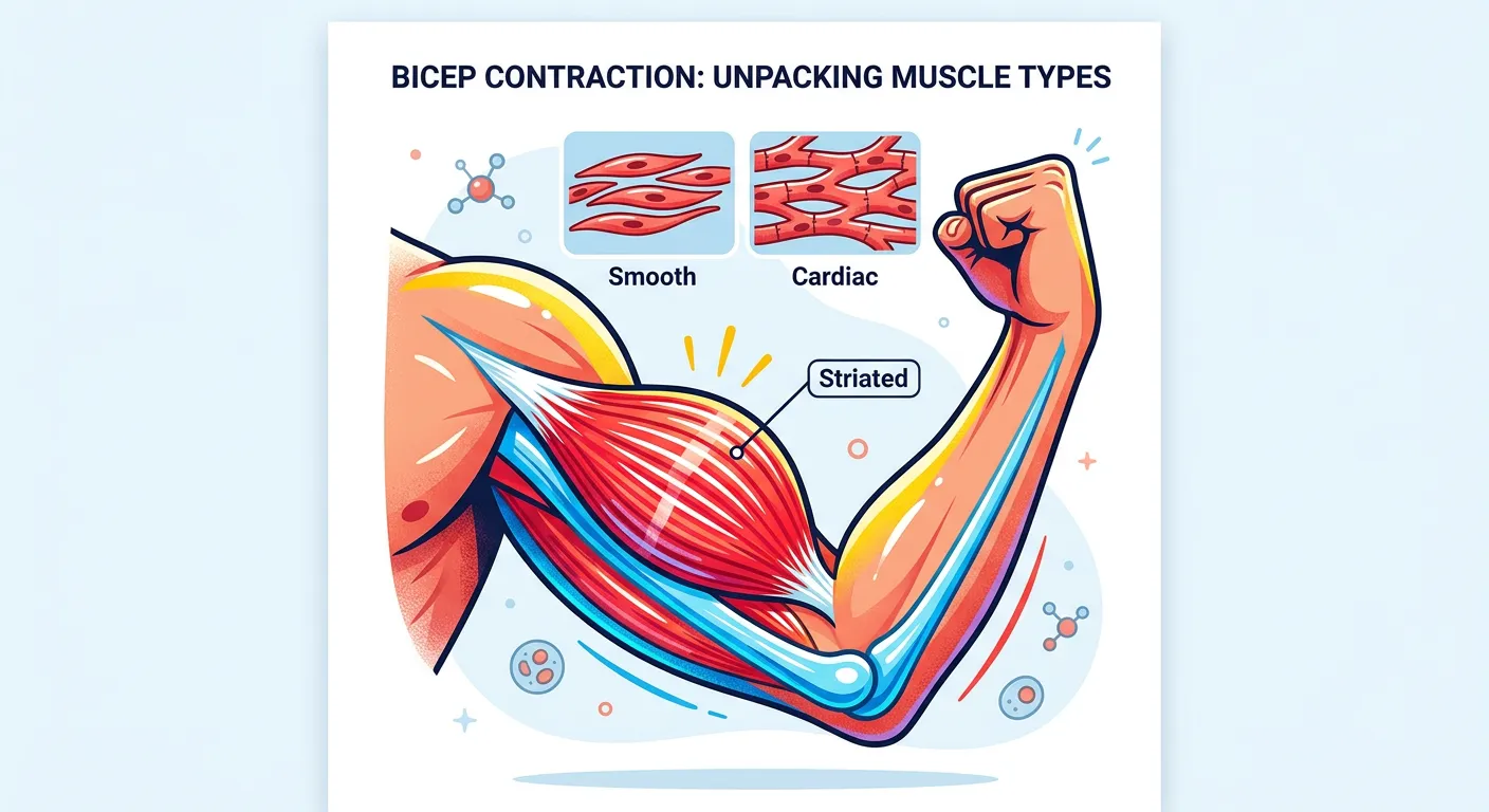
“Muscle tissue makes us move! Skeletal muscles are under our control, like lifting a bag. Smooth muscles work silently in our stomach, and Cardiac muscle is the heart's non-stop engine. They all share one superpower: the ability to contract and generate the force we need daily.”
Muscle tissue is characterized by its unique ability to contract and relax, thereby generating movement. Each muscle is made of many long, cylindrical fibers arranged in parallel arrays. These fibers are composed of even finer threads called myofibrils, which contain the contractile proteins actin and myosin. This tissue is the engine of the body, converting chemical energy from ATP into mechanical work. Without it, locomotion, the pumping of blood, and the movement of food through the gut would be impossible.
There are three distinct types of muscle tissue: Skeletal, Smooth, and Cardiac. Skeletal muscle is 'striated' (striped) and voluntary, meaning we control it consciously. These are typically attached to bones. Smooth muscle is 'non-striated' and involuntary, found in the walls of internal organs like the stomach and blood vessels. They handle slow, sustained contractions. Cardiac muscle is unique to the heart. It is striated like skeletal muscle but involuntary like smooth muscle. Its most critical feature is the 'intercalated disc,' a specialized junction that allows the heart cells to contract as a single functional unit (syncytium).
When studying for NEET, pay close attention to the structural differences. Skeletal muscles are multi-nucleated (syncytial), whereas smooth and cardiac muscles are usually uni-nucleated. The presence of striations and the level of control are the most common points of comparison used in exam questions.
| Feature | Skeletal Muscle | Smooth Muscle | Cardiac Muscle |
|---|---|---|---|
| Striations | Present | Absent | Present |
| Control | Voluntary | Involuntary | Involuntary |
| Nucleus | Multinucleated (Peripheral) | Uninucleated (Central) | Uninucleated (Central) |
| Shape | Long, Cylindrical | Spindle-shaped | Cylindrical, Branched |
| Intercalated Discs | Absent | Absent | Present |
Quick Revision Points
- Contractility: The primary property of muscle tissue facilitated by actin and myosin.
- Skeletal Muscle: Attached to bones; responsible for locomotion and posture.
- Smooth Muscle: Controls diameter of blood vessels and peristalsis in the digestive tract.
- Cardiac Muscle: Only in the heart; never fatigues; contains intercalated discs for rapid signal relay.
- Myofibrils: The contractile elements within the muscle fiber.
NEET Exam Angle
- Intercalated Discs: This is a high-yield topic. They act as communication junctions (gap junctions) allowing for coordinated heart contraction.
- Location Identification: Questions often ask which muscle type is found in the 'wall of the intestine' (Smooth) or 'biceps' (Skeletal).
- Branching Pattern: Only cardiac muscle fibers are branched. This is a key visual identifier in histological diagrams.
06Neural Tissue: Signal Propagation and the Biological Communication Network

“Neural tissue is the control center. The neuron is the star, carrying electrical signals across your body at lightning speed. It's like the internet cables in your house, ensuring your brain knows exactly what your toes are feeling. Without them, we would be completely motionless!”
Neural tissue is the body's rapid-response communication system. It exerts the greatest control over the body's responsiveness to changing conditions. The functional unit of neural tissue is the neuron. Neurons are highly specialized cells that are 'excitable,' meaning they can generate and conduct electrical impulses (action potentials) from one part of the body to another. This allows the brain to receive sensory information and send out motor commands in milliseconds.
A typical neuron consists of a cell body (Cyton), short branched projections called Dendrites (which receive signals), and a long single projection called the Axon (which sends signals). However, neurons are not the only cells in neural tissue. In fact, more than half the volume of neural tissue in our body is made up of Neuroglial cells. These 'helper' cells do not conduct impulses but instead protect, support, and insulate the neurons. Without neuroglia, neurons would be unable to function or survive.
In the NEET syllabus, neural tissue is the precursor to the 'Neural Control and Coordination' chapter. Understanding how a stimulus triggers an impulse at the neuron's membrane is fundamental. Remember that neurons are some of the longest cells in the body and, crucially, they generally do not divide once they reach maturity. This lack of regenerative capacity is why spinal cord or brain injuries are so serious.
Quick Revision Points
- Neuron Parts: Cyton (cell body), Dendrites (receivers), Axon (transmitter).
- Neuroglia: Non-excitable cells that support and protect neurons; make up >50% of neural tissue.
- Excitability: The ability of a cell to respond to a stimulus by generating an electrical impulse.
- Synapse: The functional gap between two neurons where signals are transmitted chemically.
- Origin: Neural tissue is derived from the embryonic Ectoderm.
NEET Exam Angle
- Neuroglia vs. Neurons: A common trick question involves the proportion of neuroglia. Always remember they occupy more than half the volume of neural tissue.
- Stimulus Response: Understand the sequence: Stimulus → Change in membrane potential → Nerve impulse → Signal travel along the axon.
- Diagram Labels: Be able to identify the axon, dendrites, and nodes of Ranvier on a standard neuron diagram.
07Integrating Tissue Concepts for NEET: Synthesis and Revision Strategy

“And that is the foundation of animal tissues! Remember, biology isn't just memorizing definitions; it's understanding how these tiny building blocks create the masterpiece that is you. Keep revising, stay curious, and you'll ace that NEET exam. See you in the next deep dive!”
As we conclude our deep dive into animal tissues, it is important to step back and look at the 'big picture.' Tissues do not exist for their own sake; they organize into organs like the heart, lungs, or kidneys. Each organ is usually composed of all four tissue types working in harmony. For example, the heart has an epithelial lining (endocardium), a connective tissue framework, a thick layer of cardiac muscle (myocardium), and neural tissue to regulate the heartbeat. This level of organization is what allows for the complex physiological processes we study in later units.
For your NEET preparation, the shift should now be from memorizing individual definitions to applying your knowledge to histology (the study of tissues). You must be able to look at a diagram and instantly identify the tissue based on its characteristics: Is it a single layer of flat cells? (Squamous). Is it a branched fiber with striations? (Cardiac muscle). Is it a cell with long processes? (Neuron). The most successful students are those who can link the structure of a tissue directly to the function of the organ it inhabits.
Finally, use the 'compare and contrast' method. Don't just learn about ligaments; learn them alongside tendons. Don't just learn about bone; compare it with cartilage. This approach helps eliminate confusion and prepares you for the 'Match the Following' and 'Statement-based' questions that are now frequent in the NEET exam.
| Comparison Pair | Key Differentiating Factor |
|---|---|
| Tendon vs. Ligament | Tendon: Muscle-to-Bone |
| Bone vs. Cartilage | Bone: Hard/Non-pliable |
| Axon vs. Dendrite | Axon: Away from Cyton |
| Ciliated vs. Squamous | Ciliated: Moves mucus |
Quick Revision Points
- Organ System Concept: Multiple tissues working together to perform a physiological task.
- Visual Recognition: Train your eyes to identify NCERT tissue diagrams instantly.
- Functional Linking: Always ask 'Why is this tissue here?' (e.g., Squamous in Alveoli for gas exchange).
- Revision: Focus on the 'Exceptions' and 'Unique Features' (like intercalated discs or the fluid nature of blood).
NEET Exam Angle
- Match the Following: This is the most common pattern for this topic. Connect tissue types to their specific anatomical locations.
- Statement Analysis: Be careful with statements like 'All connective tissues secrete fibers.' (False, because Blood does not).
- NCERT Focus: Stick strictly to NCERT examples, as 95% of tissue questions are derived directly from the textbook text and diagrams.
Recommended Reading
Explore related Biology topics to build deeper chapter connections for NEET.
- Morphology and Modifications · Topic 2.1
- Families · Topic 2.10
- Animal Tissues · Topic 2.11
- Frog Morphology · Topic 2.12
- Digestive System · Topic 2.13
- Circulatory System · Topic 2.14
- Jump to Key Terms (Quick Revision)
- Review Common NEET Mistakes
- Read Topic FAQs
- Check PYQ Pattern Notes
- Practice NEET MCQs
- Solve NEET PYQs
📚 Key Terms
⚠️ Common NEET Mistakes
- 1Confusing Ligaments (Bone-to-Bone) with Tendons (Muscle-to-Bone). Remember 'LBB' for Ligament-Bone-Bone.
- 2Thinking that all connective tissues secrete fibers. Remember that Blood is the major exception.
- 3Assuming that 'striated' and 'voluntary' always go together. Cardiac muscle is striated but involuntary.
- 4Misidentifying the location of ciliated epithelium. It is specific to the bronchioles and fallopian tubes in NCERT.
- 5Overlooking the fact that neuroglial cells make up more than half the volume of neural tissue.
- 6Confusing the germ layer origins. While many are single-origin, Epithelium comes from all three layers.
📝 NEET PYQ Pattern
Between 2018–2024, NEET questions have frequently focused on 'Match the Following' patterns connecting tissue types (like ciliated epithelium or tendons) to their specific anatomical locations. There is also a consistent focus on identifying tissue types from NCERT-based diagrams and understanding the unique features of cardiac muscle junctions like intercalated discs.
❓ Frequently Asked Questions
What are the main differences between skeletal, smooth, and cardiac muscles?
Skeletal muscles are striated, voluntary, and multinucleated, typically attached to bones. Smooth muscles are non-striated, involuntary, and uninucleated, found in internal organs. Cardiac muscles are striated, involuntary, branched, and contain intercalated discs for coordinated heartbeats.
Where is simple squamous epithelium located in the human body and why?
It is located in the walls of blood vessels (endothelium) and air sacs of lungs (alveoli). Its thin, single-layer structure forms a diffusion boundary, allowing for the rapid exchange of gases and nutrients.
Why is blood classified as a connective tissue despite being liquid?
Blood is classified as a connective tissue because it originates from the mesoderm and possesses an extracellular matrix (plasma) that connects all systems of the body by transporting nutrients, gases, and waste. However, unlike other connective tissues, its cells do not secrete structural fibers.
How does cuboidal epithelium assist in the functions of the kidney tubules?
In the Proximal Convoluted Tubule (PCT) of the kidney, simple cuboidal epithelium often possesses microvilli (brush border). This significantly increases the surface area for the reabsorption of essential nutrients and water from the filtrate back into the blood.
What is the structural role of collagen fibers in dense connective tissue?
Collagen fibers provide high tensile strength and structural integrity. In dense regular connective tissue like tendons, collagen fibers are oriented in parallel bundles to resist pulling forces from a specific direction.
What is the difference between specialized connective tissue and loose connective tissue?
Loose connective tissue (like areolar and adipose) has cells and fibers loosely arranged in a semi-fluid ground substance, serving as a support or storage site. Specialized connective tissues (like bone, cartilage, and blood) have unique matrices adapted for rigid support, flexibility, or fluid transport.
Written By
NEET Content Strategist & Biology Expert
Sangita Kumari is a NEET educator and content strategist with over 6 years of experience teaching Biology, Chemistry, and Physics to Class 11 and 12 aspirants. She helps bridge the gap between traditional NCERT preparation and modern AI-powered learning. Her content is trusted by thousands of NEET aspirants across India.