🎬 Video Lesson Available
Watch the full 7-slide video lesson for Cell Envelope with AI teacher narration and visual explanations.
01The Architecture of Bacterial Defense: Defining the Triple-Layered Cell Envelope
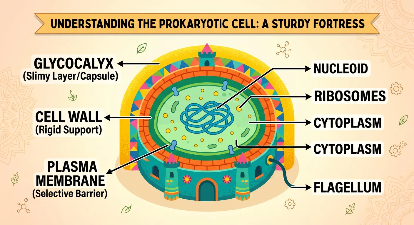
“Welcome, NEET warriors! Think of a bacteria like a medieval fort. Just as a fort has layers of defense, a bacterial cell has a 'Cell Envelope.' It is a tightly linked three-layered structure: the Glycocalyx, the Cell Wall, and the Plasma Membrane. Let's decode them!”
In the world of prokaryotes, the cell envelope is not just a simple boundary; it is a sophisticated, chemically complex protective unit. Most prokaryotic cells, especially bacteria, possess this specialized envelope that serves as their primary interface with the environment. When we look at this structure under an electron microscope, we see it isn't just one layer. Instead, it is a tightly linked, three-layered organization. While each of these three layers has a distinct chemical composition and specific biological role, they act together as a single 'Integrated Functional Unit.' This means you cannot view them in isolation; they work in tandem to ensure the cell's survival against physical and chemical stresses.
The arrangement always follows a strict hierarchy from the outermost to the innermost layer. First, we encounter the glycocalyx, followed by the cell wall, and finally the plasma membrane. For a NEET aspirant, it is crucial to understand that even though these layers are physically connected, their biochemical properties vary significantly between different species of bacteria. This variation is the basis for how we classify bacteria into two broad categories: Gram-positive and Gram-negative. This classification, developed by Christian Gram, depends entirely on how the cell envelope responds to a specific staining procedure. If the envelope retains the crystal violet stain, it is Gram-positive; if it loses it and takes up the counterstain, it is Gram-negative.
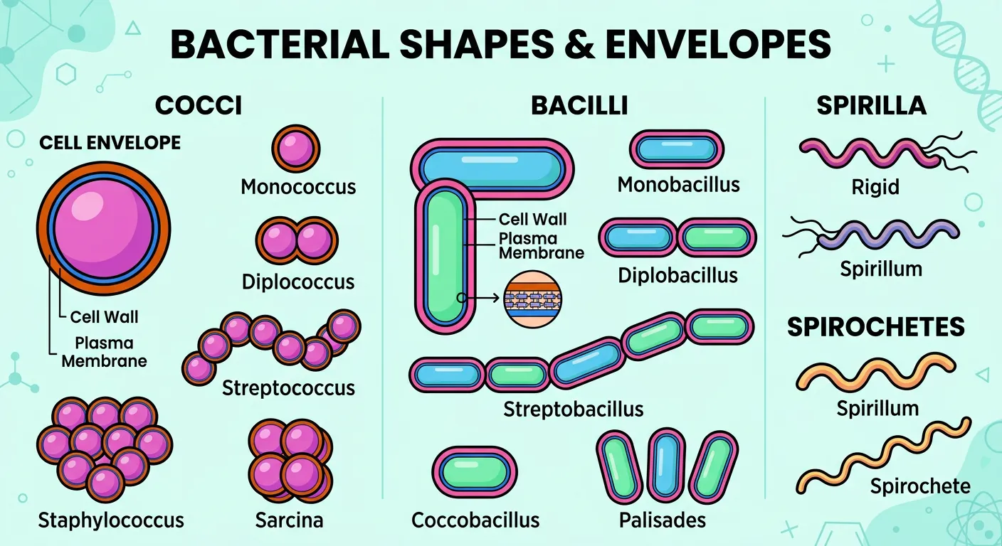
Understanding the cell envelope is the foundation of bacterial morphology. Whether a bacterium is a coccus, bacillus, or spirillum, the envelope provides the necessary structural support to maintain that shape. It is also the reason why bacteria can survive in environments that would otherwise cause a simple cell to burst or shrivel. This 'fortress' approach to cellular design is what makes bacteria some of the most resilient organisms on Earth.
Quick Revision Points
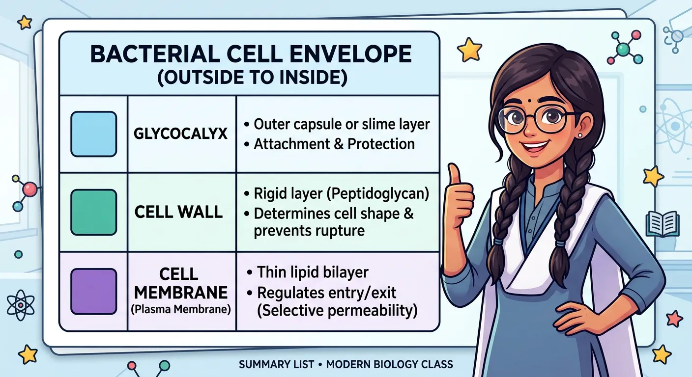
- The cell envelope is a chemically complex, three-layered protective structure.
- Layers from outside to inside: Glycocalyx → Cell Wall → Plasma Membrane.
- The three layers function together as a single Integrated Functional Unit.
- Bacteria are classified as Gram-positive or Gram-negative based on envelope differences.
- Structural integrity is maintained despite the distinct roles of each layer.
NEET Exam Angle
- NCERT Focus: Always remember the sequence of layers (Glycocalyx is outermost).
- PYQ Alert: Questions often ask about the 'Integrated Functional Unit'—this refers to the collective action of all three layers.
- Classification: Gram staining is a frequent topic; focus on the envelope's reaction to the stain.
02The Glycocalyx: Differentiating Slime Layers and Capsules in Bacterial Pathogenesis
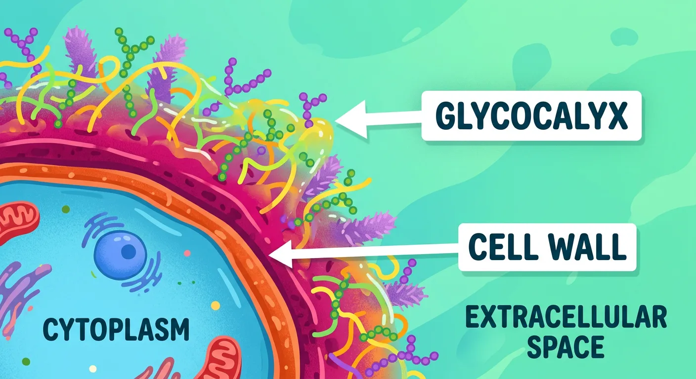
“First, the outermost layer: the Glycocalyx. Think of this like a 'sticky slime layer' that helps the bacteria stick to surfaces, or a tougher 'capsule' that protects it from our immune system's attack. It is the bacteria's first line of defense against being swallowed!”
The glycocalyx is the 'first contact' layer of the bacterial cell. Chemically, it consists of various polysaccharides and sometimes proteins, but its physical consistency varies wildly between species. In some bacteria, the glycocalyx is present as a loose, gelatinous sheath called the 'slime layer.' This layer is primary used for preventing desiccation (drying out) and helping the bacteria stick to surfaces like your teeth or medical catheters. It is relatively easy to wash away but provides a significant advantage for biofilm formation.
Conversely, in other bacteria, the glycocalyx is thick and tough, forming what we call a 'capsule.' The capsule is a major virulence factor, meaning it makes the bacteria more likely to cause disease. Why? Because a capsule acts like an 'invisibility cloak' or a shield. When pathogenic bacteria enter the human body, our immune cells (phagocytes) try to engulf and destroy them. A thick capsule prevents these phagocytes from getting a grip on the bacteria, allowing the pathogen to multiply and cause infection. This is why encapsulated strains of Streptococcus pneumoniae are deadly, while non-encapsulated strains are often harmless.
For your NEET preparation, focus on the distinction between these two forms. The chemical composition is essentially sugar-based (glyco = sugar, calyx = coat), but the structural arrangement determines the survival strategy. Whether it is a sticky slime layer for adherence or a rigid capsule for protection against the host immune system, the glycocalyx is the bacteria's primary defense against environmental and biological threats.
| Feature | Slime Layer | Capsule |
|---|---|---|
| Texture | Loose, gelatinous, and thin | Thick, tough, and rigid |
| Attachment | Loosely attached to the cell wall | Firmly attached to the cell wall |
| Function | Adhesion and prevents water loss | Protection from host immune system (phagocytosis) |
| Clinical Role | Biofilm formation on surfaces | Increases virulence (disease-causing ability) |
Quick Revision Points
- The glycocalyx is the outermost layer of the cell envelope.
- It is composed mainly of polysaccharides and occasionally polypeptides.
- A loose, thin arrangement is known as the Slime Layer.
- A thick, tough, and well-organized arrangement is called the Capsule.
- Capsules are essential for resisting phagocytosis by the host immune system.
NEET Exam Angle
- Definition Match: Match 'Capsule' with 'Thick/Tough' and 'Slime layer' with 'Loose sheath' in Column A/B questions.
- Pathogenicity: Remember that the presence of a capsule is often linked to the ability of a bacterium to cause severe disease.
- Composition: Though varying, it is predominantly a polysaccharide layer.
03The Bacterial Cell Wall: Structural Integrity and the Gram Staining Basis
“Next is the Cell Wall. It gives the cell its shape and prevents it from bursting. We categorize bacteria into Gram-positive and Gram-negative based on their wall thickness. Remember, it determines structural strength, just like the walls of a sturdy Indian home.”
Just beneath the glycocalyx lies the cell wall, the structural backbone of the prokaryotic cell. Its primary function is to provide a strong, rigid framework that determines the shape of the bacterium—whether it’s a rod (bacillus), sphere (coccus), or spiral (spirillum). Beyond aesthetics, the cell wall is a life-saver. Because bacteria often live in hypotonic environments (where the salt concentration is lower than inside the cell), water constantly tries to rush in. Without the counter-pressure of a rigid cell wall, the cell would suffer from 'osmotic lysis' and literally burst.
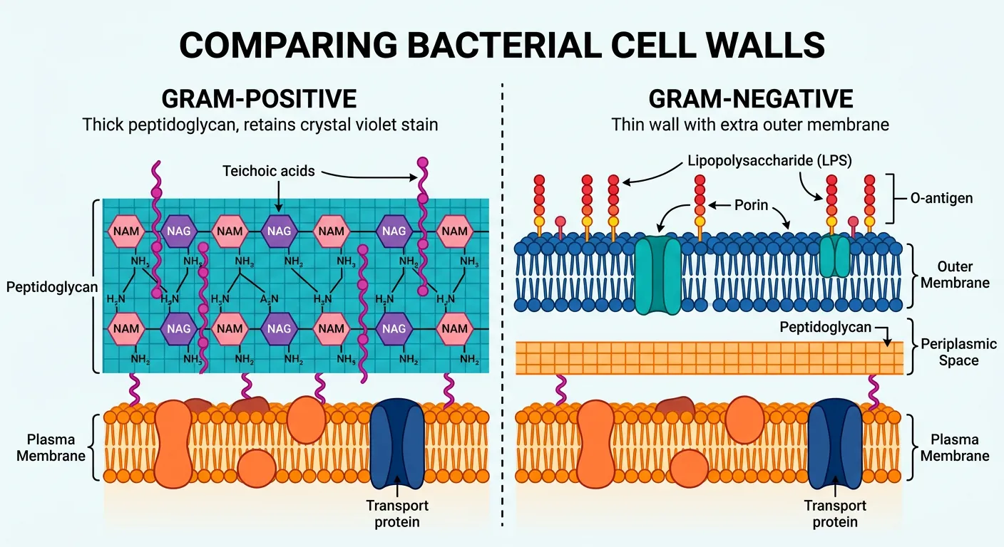
The chemical secret of the bacterial cell wall is a unique polymer called Peptidoglycan (also known as Murein). This is a mesh-like structure made of sugars and amino acids. This brings us to the famous Gram stain. Gram-positive bacteria have a very thick, multi-layered peptidoglycan wall that traps the crystal violet dye. Gram-negative bacteria, however, have a very thin layer of peptidoglycan and an extra 'outer membrane' containing lipopolysaccharides. This outer membrane makes Gram-negative bacteria more resistant to many antibiotics.
From a clinical perspective, the cell wall is the target of many common antibiotics. Penicillin, for example, works by preventing bacteria from cross-linking their peptidoglycan strands. This weakens the wall to the point where the bacteria burst and die. This is why antibiotics are so effective against bacteria but don't harm human cells (because human cells lack a cell wall entirely).
| Feature | Gram-Positive Bacteria | Gram-Negative Bacteria |
|---|---|---|
| Peptidoglycan Layer | Very thick (multiple layers) | Very thin (single layer) |
| Outer Membrane | Absent | Present |
| Lipopolysaccharides | Virtually absent | High content |
| Stain Color | Purple / Violet | Pink / Red |
| Antibiotic Sensitivity | Generally more susceptible | Generally more resistant |
Quick Revision Points
- The cell wall determines the cell shape and provides structural support.
- It prevents the bacterium from bursting (lysis) in hypotonic solutions.
- Peptidoglycan is the primary chemical component unique to bacteria.
- Gram-positive cells have thick walls; Gram-negative cells have thin walls plus an outer membrane.
- The cell wall is a primary target for antibiotics like Penicillin.
NEET Exam Angle
- Mechanism: Focus on how the cell wall prevents osmotic lysis (a frequent conceptual question).
- Differences: Be clear on the Gram-positive vs. Gram-negative wall structure.
- Composition: Peptidoglycan (Murein) is the keyword to remember for bacterial walls.
04The Plasma Membrane: Selective Permeability and the Gatekeeper of Metabolism
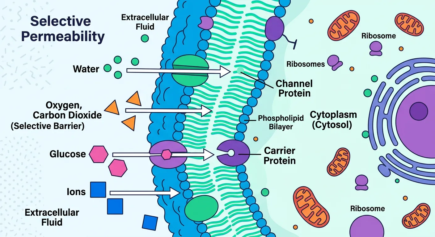
“Deep inside lies the Plasma Membrane. It is semi-permeable, acting like a strict security guard at a gate. It decides what enters and exits the cell, maintaining the internal environment. It is the ultimate decision-maker for the cell's metabolic activity.”
The innermost layer of the cell envelope is the plasma membrane. While the cell wall is rigid and protective, the plasma membrane is fluid and functional. In prokaryotes, the plasma membrane is semi-permeable in nature, meaning it acts as a selective barrier. It controls exactly what enters the cell (nutrients, ions) and what exits (waste products, secreted proteins). It interacts directly with the outside world, making it the 'sensory organ' of the cell.
One of the most fascinating points for NEET is the evolutionary conservation of the plasma membrane. Chemically, the prokaryotic plasma membrane is remarkably similar to the eukaryotic plasma membrane. Both consist of a phospholipid bilayer with embedded proteins. However, there is a key difference you must remember: while eukaryotic membranes contain sterols (like cholesterol) to provide stability, most prokaryotic membranes (except for Mycoplasma) lack sterols. Instead, they often use hopanoids, which are sterol-like molecules, to maintain membrane fluidity.
In prokaryotes, the plasma membrane takes on many jobs that organelles do in eukaryotes. Since bacteria lack mitochondria, the enzymes for the Electron Transport Chain (ETC) and ATP production are actually embedded directly into the plasma membrane. It isn't just a container; it's the metabolic powerhouse of the cell. It also plays a role in sensing environmental changes—like pH or temperature—and triggering the cell to move toward food or away from toxins (chemotaxis).
Quick Revision Points
- The plasma membrane is the innermost component of the cell envelope.
- It is semi-permeable, regulating the traffic of molecules in and out.
- Structurally similar to eukaryotic membranes (phospholipid bilayer).
- Most prokaryotic membranes lack sterols (unlike eukaryotes).
- It hosts vital metabolic processes like respiration and sensing environmental signals.
NEET Exam Angle
- Comparison: NEET often asks about the similarity between prokaryotic and eukaryotic membranes. Focus on the phospholipid bilayer.
- Exception: Mycoplasma is a rare prokaryote that has sterols in its membrane and lacks a cell wall.
- Role: Remember that the plasma membrane in bacteria performs functions of the mitochondria (respiration).
05Mesosomes and Membrane Extensions: Specialized Prokaryotic Internal Workshops
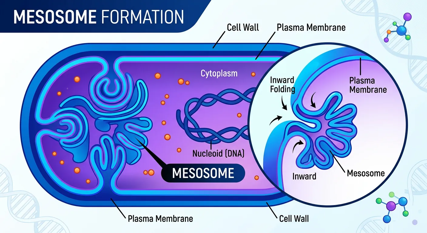
“Look closely at the membrane! Sometimes it folds into special structures called Mesosomes. These are like tiny, folded internal workshops that help in respiration, DNA replication, and secretion. They are the 'powerhouse' regions for prokaryotes, performing functions similar to our mitochondria!”
Prokaryotes may lack membrane-bound organelles, but they have a clever workaround: mesosomes. Mesosomes are specialized membranous structures formed by the extension or 'infolding' of the plasma membrane into the cell. They can appear in three physical forms: vesicles (bag-like), tubules (tube-like), or lamellae (sheet-like). These aren't just random folds; they are highly organized regions of activity that compensate for the lack of complex internal compartments.
The functions of mesosomes are diverse and essential for the cell cycle. They play a critical role in cell wall formation by organizing the necessary enzymes. During cell division, they assist in DNA replication and the equal distribution of the replicated DNA to daughter cells. By increasing the surface area of the plasma membrane, mesosomes also increase the space available for enzymatic activity and respiratory processes. This is why they are often called the 'prokaryotic analog to mitochondria.'
Beyond mesosomes, some prokaryotes, like Cyanobacteria (blue-green algae), have other membrane extensions called 'chromatophores.' These contain pigments like chlorophyll and are responsible for photosynthesis. For a NEET student, it is vital to distinguish between these two: Mesosomes are for respiration and replication, while Chromatophores are for photosynthesis. Both, however, are proof of the plasma membrane's incredible versatility in prokaryotic life.
| Form of Mesosome | Description | Primary Association |
|---|---|---|
| Vesicles | Small, sac-like structures | Secretion and storage |
| Tubules | Long, tube-like extensions | Increasing surface area for enzymes |
| Lamellae | Layered or sheet-like folds | Respiration and photosynthesis (in some) |
| Functions | DNA Replication, Cell Wall synthesis, Respiration | General cellular maintenance |
Quick Revision Points
- Mesosomes are internal folds of the plasma membrane in prokaryotes.
- They appear as vesicles, tubules, or lamellae.
- Functions: Cell wall synthesis, DNA replication, and secretion processes.
- They increase surface area for respiration and enzymatic actions.
- Chromatophores are similar extensions in Cyanobacteria containing photosynthetic pigments.
NEET Exam Angle
- Surface Area: The most common answer for 'Why do mesosomes exist?' is to increase surface area for enzymatic/respiratory activity.
- DNA Distribution: Remember mesosomes help in distributing DNA to daughter cells during binary fission.
- Analogy: Mesosomes are to Prokaryotes what Mitochondria are to Eukaryotes.
06Survival Strategies: Evolutionary Modifications of the Cell Envelope
“The cell envelope is not just a boundary; it is a specialized survival kit. Depending on the species, these layers undergo modifications to help the bacteria survive in extreme environments, whether it is high heat or acidic conditions. Evolution at its finest!”
The cell envelope is the ultimate tool for bacterial adaptation. Evolution has modified these three layers to allow bacteria to thrive in the most hostile environments on Earth. For example, Archaebacteria (the most ancient of prokaryotes) often live in extreme heat (thermophiles) or high salt (halophiles). Their cell envelopes differ significantly from standard bacteria; their membranes contain branched-chain lipids which provide extra stability against heat that would melt a normal cell membrane.
In photosynthetic bacteria like Cyanobacteria, the envelope modifications are even more specialized. These organisms developed chromatophores—membranous extensions containing pigments—to capture sunlight. This was a massive evolutionary leap, allowing prokaryotes to become self-sufficient producers. Furthermore, many bacteria produce 'appendages' that are extensions of the cell envelope. Flagella (for motility), Pili (for genetic exchange), and Fimbriae (for attachment) are all anchored into the envelope layers. While these aren't part of the 'triple layer' itself, they are critical modifications for survival.
Finally, consider the medical importance of these modifications. Some bacteria can shed their cell wall temporarily to become 'L-forms,' making them invisible to antibiotics that target the cell wall. Others use their envelope to pump out toxins or antibiotics (efflux pumps). Understanding how the cell envelope modifies itself is not just a biology lesson; it's the key to understanding how life persists in extreme niches and how pathogens evolve resistance to our medicines.
| Modification | Organism Example | Primary Survival Benefit |
|---|---|---|
| Branched Lipids | Archaebacteria | Heat and acid resistance |
| Chromatophores | Cyanobacteria | Photosynthetic pigment storage |
| Fimbriae | E. coli | Adhesion to host tissues |
| Efflux Pumps | Drug-resistant bacteria | Pumping out harmful antibiotics |
Quick Revision Points
- Cell envelope modifications allow survival in extreme environments (heat, salt, acid).
- Archaebacteria use branched-chain lipids for membrane stability.
- Chromatophores are specialized pigment-containing extensions in Cyanobacteria.
- Appendages like flagella, pili, and fimbriae are anchored in the cell envelope.
- The envelope is dynamic and can change to resist environmental or clinical stress.
NEET Exam Angle
- Archaebacteria: Note that their membrane structure is a key reason they can survive extreme conditions.
- Cyanobacteria: Distinguish between Mesosomes (general) and Chromatophores (pigmented).
- Motility: Remember that flagella are extensions of the cell envelope, not just the cell wall.
07NEET Summary and High-Yield Revision: Mastering Bacterial Structures
“So, remember the trio: Glycocalyx for protection, Cell Wall for shape, and Plasma Membrane for control. Master this envelope, and you have secured marks on bacterial structure! Keep studying hard, stay focused, and keep pushing toward your MBBS dream. See you in the next lecture!”
As we wrap up our deep dive into the prokaryotic cell envelope, let’s consolidate the 'must-know' facts for your NEET exam. The concept of the 'Integrated Functional Unit' is your starting point—never forget that the glycocalyx, cell wall, and plasma membrane work as a team. If a question asks about the order, it's always Glycocalyx (out) → Cell Wall (middle) → Plasma Membrane (in). If it asks about classification, immediately think of the Gram stain and the thickness of the peptidoglycan layer.
Focus your revision on the functional differences between the forms of the glycocalyx. A capsule is thick and tough for protection (virulence), while a slime layer is loose for adherence and moisture retention. In the cell wall section, focus on its role in preventing osmotic lysis. For the plasma membrane, remember its semi-permeable nature and its specialized infoldings: the mesosomes. Mesosomes are perhaps the most frequently tested sub-topic here; remember they help in respiration, DNA replication, and increasing surface area.
Lastly, don't confuse the various extensions. Flagella are for movement, pili are for 'mating' (conjugation), and fimbriae are for sticking to things. Chromatophores are for photosynthesis and are exclusive to certain bacteria like cyanobacteria. Mastering these distinctions will ensure you can navigate any 'match the column' or 'statement-based' question that NEET throws at you. You are now equipped with the knowledge to conquer this topic. Keep your NCERT handy, practice the diagrams, and stay consistent in your revision. Your seat in medical college is waiting!
Quick Revision Points
- Trio functions: Glycocalyx (Protection), Cell Wall (Shape/Strength), Plasma Membrane (Control/Metabolism).
- Gram Stain: Gram-positive (Purple, thick wall); Gram-negative (Pink, thin wall + outer membrane).
- Mesosomes: Infoldings for respiration and DNA replication; increase surface area.
- Pathogenicity: Capsules help bacteria evade the host's immune system.
- Osmotic Protection: The cell wall is vital to prevent the cell from bursting in water.
NEET Exam Angle
- Exam Pitfall: Don't confuse 'capsule' with 'cell wall'; the capsule is outside the wall.
- Statement Questions: Watch out for statements saying prokaryotic membranes are completely different from eukaryotic ones (they are actually quite similar).
- Focus Areas: Mesosomes and the differences in Gram staining are high-probability topics.
Recommended Reading
Explore related Biology topics to build deeper chapter connections for NEET.
- Cell Theory · Topic 3.1
- Golgi Bodies · Topic 3.10
- Lysosomes · Topic 3.11
- Vacuoles · Topic 3.12
- Plastids · Topic 3.15
- Prokaryotic and Eukaryotic Cell · Topic 3.2
- Jump to Key Terms (Quick Revision)
- Review Common NEET Mistakes
- Read Topic FAQs
- Check PYQ Pattern Notes
- Practice NEET MCQs
- Solve NEET PYQs
📚 Key Terms
⚠️ Common NEET Mistakes
- 1Thinking the capsule is the same as the cell wall; remember the capsule is an additional outer layer.
- 2Confusing Gram-positive with pink color; remember Gram-Positive is Purple/Violet.
- 3Assuming all bacteria have mesosomes for photosynthesis; mesosomes are for respiration, while chromatophores are for photosynthesis.
- 4Believing that the plasma membrane of prokaryotes is identical to eukaryotes; remember prokaryotes usually lack sterols.
- 5Mixing up the functions of fimbriae and flagella; flagella are for movement, while fimbriae are for attachment.
📝 NEET PYQ Pattern
Recent NEET papers (2018–2024) show a high frequency of questions on the functions of mesosomes and the hierarchical layers of the cell envelope. Matching-type questions regarding the types of glycocalyx (Capsule vs. Slime layer) and statement-based questions on the Gram stain mechanism are very common. Pay special attention to the role of mesosomes in DNA distribution and surface area enhancement.
❓ Frequently Asked Questions
What are the three components of the bacterial cell envelope?
The bacterial cell envelope consists of three layers: the outermost glycocalyx, the middle cell wall, and the innermost plasma membrane. Together, they act as a single integrated functional unit.
How does a bacterial capsule differ from a slime layer in function and structure?
A capsule is a thick, tough, and organized layer that firmly attaches to the cell wall, primarily helping bacteria evade the host immune system. A slime layer is a thin, loose, and gelatinous sheath that helps in attachment to surfaces and prevents water loss.
What role do mesosomes play in prokaryotic cell respiration and DNA replication?
Mesosomes increase the surface area of the plasma membrane, providing more space for respiratory enzymes. They also assist in cell wall synthesis, DNA replication, and the distribution of chromosomes to daughter cells during division.
Why is the plasma membrane of bacteria called 'semi-permeable'?
It is called semi-permeable because it selectively allows certain molecules (like nutrients and ions) to pass through while blocking others, thereby maintaining the internal environment of the cell.
What is the primary chemical difference between Gram-positive and Gram-negative cell walls?
Gram-positive cell walls have a very thick layer of peptidoglycan. Gram-negative cell walls have a much thinner layer of peptidoglycan but possess an additional lipid-rich 'outer membrane'.
How does the cell wall prevent a bacterial cell from bursting in a hypotonic environment?
The cell wall is rigid and provides structural strength. When water enters the cell via osmosis, the cell wall exerts counter-pressure, preventing the plasma membrane from expanding to the point of rupture (osmotic lysis).
Written By
NEET Content Strategist & Biology Expert
Sangita Kumari is a NEET educator and content strategist with over 6 years of experience teaching Biology, Chemistry, and Physics to Class 11 and 12 aspirants. She helps bridge the gap between traditional NCERT preparation and modern AI-powered learning. Her content is trusted by thousands of NEET aspirants across India.